

We present the case of a 65-year-old male patient, phenotype negative carrier of a pathogenic variant in the desmoglein-2 gene (DSG2), with a normal cardiac magnetic resonance imaging (CMR).
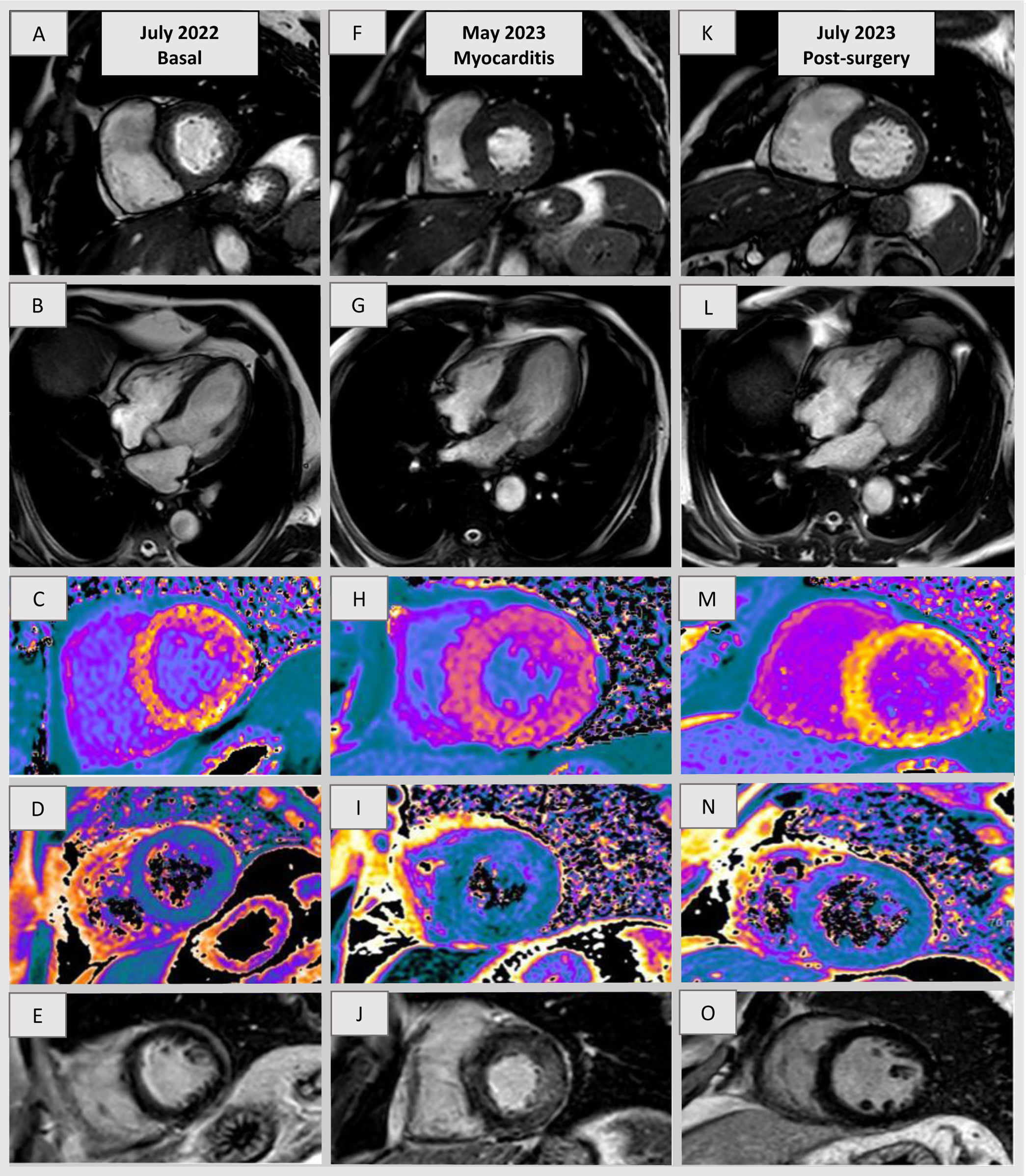
He was admitted to hospital with the initial diagnosis of acute myocardial infarction without ST-segment elevation, hypertension and tachycardia. Coronary angiography showed no alterations. CMR revealed prolonged T1 mapping (Fig. 1H), T2 mapping, increased extracellular volume (Fig. 1I), and the presence of late gadolinium enhancement with diffuse hyperintensity, predominantly in the midwall, and lateral and anterobasal segments (Fig. 1J), fulfilling Lake Louise criteria for myocarditis. Left ventricle hypertrophy and systolic anterior motion of the anterior mitral leaflet were also patent (Fig. 1F, G).
The myocarditis episode was initially attributed to SARS-CoV-2 infection. During follow-up, the patient reported intermittent episodes of headache, facial flushing, sweating, and hypertension, suspicious of pheochromocytoma symptoms, which was confirmed after positive urine metanephrine test and abdominal computed tomography, revealing a 40mm left adrenal mass that was later confirmed by pathological examination. The patient underwent surgical intervention with no complications. Subsequent CMR demonstrated complete regression of left ventricle hypertrophy (Fig. 1K, L), edema (Fig. 1N) and late gadolinium enhancement (Fig. 1O). Notably, the left ventricle wall thickness reduced from 17 to 11mm and the T1 mapping value was reduced from 1205ms to 1009ms and T2 mapping value reduced from 60ms to 48ms. The figure shows the basal CMR (Fig. 1A–E), during myocarditis episode (Fig. 1F–J) and post-surgery (Fig. 1K–O).
FundingNo funding sources were required for this work.
Ethical considerationsThe authors confirm that informed consent was obtained from the patient for the dissemination of the case. We confirm that the study adheres to the Sex and Gender Equity in Research (SAGER) guidelines, ensuring the consideration of sex and gender dimensions throughout the research process.
Statement on the use of artificial intelligenceWe confirm that no artificial intelligence (AI) tools were used in the design, conduct, analysis, or reporting of this study. All work was carried out by the authors without the assistance of AI technologies.
Authors’ contributionsJ. Zubiaur participated in the drafting, preparation and correction of the manuscript. L. Ruiz Guerrero, G. Martín Gorria and V. Fernández Lobo participated in the drafting and revision of the manuscript.
Conflicts of interestNone.