


A 59-year-old woman with history of hypertension, dyslipidemia and a well differentiated gastric neuroendrocrine tumor on stage IV due to hepatic metastases, under somatostatin analogs treatment, was admitted at our center due to increasing shortness of breath, orthopnea, and peripheral edema. On physical examination, she was tachypneic, her oxygen saturation was 88% and had a blood pressure of 170/80. Cardiac auscultation revealed a 4/6 diastolic murmur along the left sternal border. The chest X-ray showed pleural effusion and a NTproBNP of 16.400pg/mL was remarkable in laboratory results. The transthoracic echocardiography revealed enlarged left ventricle with mild dysfunction. Severe aortic regurgitation and mild anterior leaflet prolapse with moderate to severe mitral regurgitation was exposed. The transesophageal echocardiography, in parasternal short-axis view of the great vessels showed a quadricuspid aortic valve (QAV) with the presence of 3 equal sized thin cusps and a minor one, with a “X-shaped” commissural aspect in diastole (Fig. 1). There was a severe (grade 4) central aortic regurgitation, resulting from an incomplete diastolic coaptation of the cusps. The aortic root and the ascending aorta were not dilated. The coronary angiography was normal. Due to refractory heart failure despite medical treatment with diuretics and intravenous nitroglycerine, and considering long life expectancy by our Oncology team, she was promptly operated. The prosthetic aortic valve was implanted and mitral valvuloplasty was uneventfully performed. After a long recovery, she remains in New York Heart Association class I 1 year after the operation.

Transesophageal echocardiogram. A, B: quadricuspid aortic valve with the presence of 3 equal sized thin cusps and a minor one. C, D: color Doppler reveals severe aortic regurgitation due to lack of central coaptation. E, F: anterior leaflet mitral valve prolapse causing moderate mitral regurgitation.
The QAV is a rare manifestation of congenital aortic valve abnormalities, with an incidence of 0.008% at autopsy and 1% in patients presented for AV surgery.1 QAV anatomy is variable, and according to this, Hurwitz and Roberts2 categorized QAV type into 7 subtypes (A to G). The 2 most frequent types are type A (4 equal cusps) and type B (3 normal cusps with 1 smaller cusp). Abnormal cusps may be formed as a result of a developmental anomaly during embryological arterial trunk septation, although the mechanism of this congenital malformation is not fully known. One of the leading hypotheses is abnormal septation of embryological truncus arteriosus, with the fourth cusp arising during the early stage of truncal septation resulting to either a different number of primordial aortic leaflets or an abnormal cusp proliferation.
The functional aspect of the quadricuspid valve is mainly represented by pure insufficiency; aortic stenosis may be present but is rare. Anatomical abnormalities of the cusps could induce unequal shear stress leading to fibrosis and incomplete coaptation so that it is thought that type B has a greater probability of developing aortic regurgitation because of the asymmetric shear stress of the cusps. Valve replacement is often required by the fifth or sixth decade of life.1
Some QAVs are often associated with other abnormalities, such as displacement of the coronary sinus and ostium, ventricular septal defect, patent duct arteriosus, subaortic stenosis, hypertrophic cardiomyopathy, and Valsalva aneurysm.
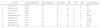
Mitral valve prolapse (MVP) is a common cause of mitral regurgitation, and primary MVP can be associated with other valvular abnormalities (tricuspid valve in up to 40 to 50 percent of patients, and aortic valve in 10 to 20 percent), usually as a prolapse3; or with the most common congenital aortic valvular anomaly, the bicuspid valve. However, few cases of QAV and MVP have been published4 (Table 1).
Reported cases of coexistence of quadricuspid aortic valve and mitral valve prolapse.
| Author | Year of publication | Age of diagnosis | QAV type | AR | AS | MR | Valve replaced | |
|---|---|---|---|---|---|---|---|---|
| 1 | Cooke et al. | 2000 | 75 | A | 1+ | 0 | 4+ | MV |
| 2 | Yildrim et al. | 2008 | 10 | B | 4+ | 4+ | 0 | AV |
| 3 | Di Pino et al. | 2008 | 11 | B | 0 | 0 | 1+ | None |
| 4 | Ozlü et al. | 2008 | 72 | A | 3+ | 0 | 4+ | MV |
| 5 | Jagannath et al. | 2011 | 75 | C | 3+ | 0 | 3+ | None |
| 6 | George et al. | 2013 | 41 | A | 1+ | 0 | 4+ | None |
| 7 | George et al. | 2013 | 18 | A | 1+ | 0 | 1+ | None |
| 8 | George et al. | 2013 | 54 | B | 1+ | 0 | + | None |
| 9 | Reported case | 2018 | 59 | B | 4+ | 0 | 3+ | Both |
AS, aortic stenosis; AR, aortic regurgitation; AV, aortic valve; MR, mitral regurgitation; MV, mitral valve; QAV, quadricuspid aortic valve.
QAV type. A: Four equal cusps. B: Three equal cusps and one smaller cusp. C: Two equal larger cusps and two equal smaller cusps.
Valvular dysfunction scale: 0=none, 1+=mild, 2+=mild to moderate, 3+=moderate, +=severe.
References are available in the supplementary data.
The occurrence of both in a series of patients supports the view that MVP and QAV may be associated with congenital anomalies.5 A genetic cause has even been hypothesized, as QAV has been reported in a 16-year-old boy with a clinical diagnosis of Ehlers–Danlos disease caused by a homozygous TNXB defect, in which MVP has been reported, and MVP in his mother.6
Although previous review demonstrated a more benign course of the aortic valvulopathy, with no patients requiring surgery of both valves,4 our case agrees with the tendency of QAV function to deteriorate described in the literature, probably related to the older age of our patient.
The coexistence of both mitral and aortic regurgitation in a patient with MVP requires meticulous evaluation of the aortic valve anatomy to rule out bicuspid or quadricuspid valves. Early detection of these congenital abnormalities is essential for the prevention of valvular and ventricular dysfunction and close follow up of these patients is mandatory to avoid complications.
The following are the supplementary data to this article:


