A 65-year-old man presented to the emergency department with progressive asthenia. One year ago, he noted the onset of exertional dyspnea without a history of weight loss, fever or night sweating. The initial 12-lead electrocardiogram indicated typical atrial flutter with 2:1 atrioventricular conduction. On physical examination, he was found to have elevated jugular venous pressure without bibasal rales. No lymphadenopathy was detected.
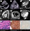
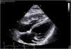
Transthoracic echocardiography revealed a severe thickening (20 mm) right ventricle free wall (Fig. 1A,B), video 1 of the supplementary data – subcostal view) with a severe right ventricular systolic dysfunction. Minimal pericardial effusion with mild pericardial thickening was detected.

Cardiac magnetic resonance imaging characterized a mass with no evidence of necrosis which infiltrated the epicardial adipose tissue as well as the right ventricle free wall with extension to the wall of the right atrium (Fig. 1C–E, and video 2 of the supplementary data). Severe right ventricular systolic dysfunction was confirmed. A thoracic and abdominal CT scan was performed (Fig. 1F) as well as a bone marrow biopsy which ruled out neither extension nor origin of extracardiac malignancy.
Final diagnosis of primary cardiac diffuse B-cell lymphoma (Fig. 1G-I) was achieved with an echo-guided intravascular biopsy of the right venricle free wall at the cath lab. Treatment with cyclofosfamide bolus (750mg) and metilprednisolone was initiated. Exitus laetalis occurred 3 months after discharge due to progression of the primary cardiac lymphoma.