

Suggestions
Idioma
metricas
Publish in this journal
Guide for authors
Searcher
Journal Information
Vol. 60. Issue 3.
Pages 161-238 (July - September 2025)
Cite
Cite
Share
Download PDF
More article options
Vol. 60. Issue 3.
Pages 161-238 (July - September 2025)
Editorial
Full text access
Sofosbuvir and QT interval: A new chapter in the safety profile of direct-acting antivirals
Sofosbuvir e intervalo QT. Un nuevo capítulo en el perfil de seguridad de los antivirales de acción directa
Visits
1250
Carlos Minguito Carazoa,b,c, Moisés Rodríguez Mañeroa,b,c,
Corresponding author
moirmanero@gmail.com

Corresponding author.
a Departamento de Cardiología, Hospital Universitario Santiago de Compostela, Santiago de Compostela, A Coruña, Spain
b Instituto de Investigación Sanitaria (IDIS), Universidad de Santiago de Compostela, Santiago de Compostela, A Coruña, Spain
c Centro de Investigación Biomédica en Red de Enfermedades Cardiovasculares (CIBERCV), Instituto de Salud Carlos III, Madrid, Spain
Related content
Olga Neva López-García, Daniel García-Arribas, Carmen Olmos, María José Devesa Medina, Javier Higueras Nafría, Sonia Izquierdo Rubio, Francisca Cuenca Alarcón, Maite Maroto Castellanos, Ana Fernández-Vega, Alejandro Cruz-Utrilla, Pablo Martínez-Vives, Enrique Rey
This item has received
Article information
Full Text
Bibliography
Download PDF
Statistics
Figures (1)
fig0005
Full Text

In the last decade, the advent of direct-acting antivirals (DAA) has dramatically changed the landscape of hepatitis C virus (HCV) treatment. With sustained virologic response (SVR) rates surpassing 95%, shorter treatment durations, and minimal side effects, DAAs—particularly sofosbuvir—have revolutionized management strategies.1 The clinical success of DAAs has also helped reduce liver-related morbidity and mortality, shifting HCV management from a specialized to a more general practice domain. However, as their use expands into broader populations, including those with advanced age, multiple comorbidities, and polypharmacy, careful evaluation of their cardiovascular safety profile becomes essential.

In a recent paper published in REC: CardioClinics, López-García et al. present a prospective observational study that explores the electrocardiographic impact of sofosbuvir and other DAAs on cardiac conduction in real-world HCV-infected patients.2 This multicenter work provides valuable insight into a potentially under-recognized adverse effect: QTc interval prolongation. The study addresses a gap in literature by providing prospective ECG assessments in a relatively unselected population initiating antiviral therapy in routine clinical practice.

The study followed 101 patients initiating antiviral therapy with or without sofosbuvir. Each patient underwent three electrocardiograms: at baseline, during treatment (week 4), and after treatment completion. The authors found that sofosbuvir was associated with a modest but statistically significant QTc prolongation at week 4 (mean increase of 5.5ms), which normalized after treatment discontinuation. Importantly, this effect was not observed in patients receiving other DAA regimens without sofosbuvir. The transient nature of this prolongation and its resolution after treatment completion suggest a drug-related but reversible mechanism.

These findings are not without precedent. Since the COVID-19 pandemic, attention has increasingly focused on the potential QTc-prolonging effects of certain antiviral agents, as well as on the best strategies for electrocardiographic monitoring in these patients.3,4 Regarding DAA for HCV, earlier pharmacovigilance reports, including alerts from the United States Food and Drug Administration (FDA), linked sofosbuvir—especially in combination with amiodarone—to serious bradyarrhythmias, including severe sinus pauses needing pacemaker implantation.5,6 Although such interactions are now well-recognized and largely avoidable, the present study suggests that sofosbuvir may exert direct or indirect effects on ventricular repolarization even in the absence of amiodarone or other bradycardic agents like in this study (none of the patients received a class III antiarrhythmic drug).

The pathophysiological basis of this effect remains elusive. Several mechanisms have been proposed to explain sofosbuvir's effects on cardiac electrophysiology, particularly when co-administered with amiodarone. The combination of sofosbuvir and amiodarone can lead to severe and symptomatic bradycardia. Studies have shown that sofosbuvir enhances the amiodarone-induced disruption of calcium handling in cardiomyocytes, which may result in alterations in cellular electrophysiology and mechanical activity. These include a pharmacodynamic interaction that leads to enhanced bradycardic effects, likely due to impaired calcium handling in cardiomyocytes, as shown in human induced pluripotent stem cell (hiPSC)-derived models.7 Additionally, structural studies have demonstrated that sofosbuvir, in the presence of amiodarone, can anchor within the pore domain of L-type calcium channels, blocking ion flow and potentiating channel inhibition.8 These findings support the hypothesis that sofosbuvir may influence cardiac repolarization through both functional and structural modulation of calcium channel activity, especially under certain pharmacologic conditions. Moreover, sofosbuvir is metabolized in the liver and excreted renally, which raises the possibility that accumulation of its metabolites in patients with hepatic or renal impairment might unmask electrophysiological effects not seen in healthier individuals. However, clinical data have been inconsistent. For instance, some studies reported no significant QTc changes in similar cohorts,9 although differences in ECG timing, measurement methods, and patient characteristics may explain the discrepancy.

From a clinical standpoint, the key question is whether this QTc prolongation carries real-world implications. In the current study, 4 (6.6%) male patients reached pathological QTc thresholds (>470ms) during therapy, though without documented arrhythmic events. Only 1 patient experienced non-sustained ventricular tachycardia (NSVT) and frequent premature ventricular contractions, findings that were not further investigated. As such, the clinical impact remains uncertain. Nevertheless, the absence of overt arrhythmic events should not lead to complacency, especially considering that QTc prolongation is a well-established surrogate marker for proarrhythmic risk.

One notable strength of this study is its real-world design, incorporating a diverse group of patients with varying degrees of liver fibrosis and comorbidities. Interestingly, the authors observed a more pronounced QTc increase in patients not receiving betablockers and in those taking other QT-prolonging medications. These findings are clinically relevant, as they highlight the potential for additive or synergistic effects on cardiac repolarization in polypharmacy settings. It also suggests that betablockers may exert a protective effect by reducing sympathetic tone, although this hypothesis requires further exploration.

The authors’ recommendation for increased vigilance—particularly ECG monitoring at baseline and during the first month of treatment—seems prudent, especially in patients with other risk factors such as advanced liver fibrosis, structural heart disease, or concomitant use of QT-prolonging drugs. In high-risk individuals, additional monitoring strategies such as Holter monitoring or telemetry may be justified, although cost-effectiveness considerations remain.

This study also raises broader questions about regulatory labeling and pharmacovigilance. Currently, the main warning related to sofosbuvir involves its combination with amiodarone. However, should this warning be extended to other populations? Would it be advisable to incorporate routine QTc monitoring into the treatment algorithm for specific subgroups? The present findings suggest that more nuanced guidance may be warranted. Furthermore, inclusion of QTc-related data in future randomized trials and post-marketing surveillance efforts could help establish clearer safety thresholds.

Several limitations must be acknowledged. The QTc was measured manually rather than using automated software, which may introduce inter-observer variability. Continuous rhythm monitoring was not employed, potentially missing transient or asymptomatic arrhythmic events. The sample size, while respectable, limits the ability to detect rare but clinically significant events such as torsades de pointes. Another important point in this study is that there were more patients taking other QTc-prolonging medications in the sofosbuvir group (24.6%) compared to those receiving other therapeutic regimens (7.5%). This could partially explain the minimal QTc interval prolongation observed in patients treated with sofosbuvir. Finally, the diversity of DAA regimens and the real-life nature of the study, while adding external validity, also introduce heterogeneity that complicates interpretation.

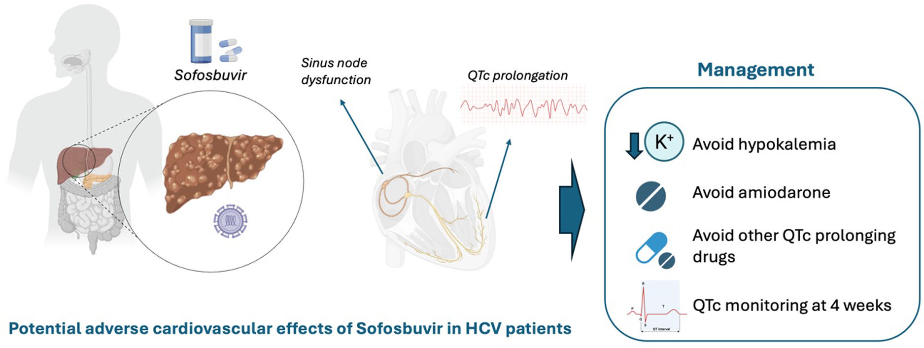
In conclusion, this study contributes to a growing body of evidence suggesting that sofosbuvir, though highly effective and generally well-tolerated, may not be entirely inert from a cardiac electrophysiology standpoint. While the observed QTc prolongation appears reversible and clinically silent in most cases, caution is warranted in specific patient subgroups. Clinicians should maintain a high index of suspicion and consider ECG monitoring in patients with risk factors for QTc prolongation. Based on the study published by López-García et al. and the existing evidence, we propose that QTc interval monitoring in patients scheduled to receive treatment with sofosbuvir should be performed at 4 weeks after treatment initiation (Fig. 1).

Fig. 1.

Proposed clinical algorithm for QT interval monitoring in patients referred for treatment with sofosbuvir. HCV, hepatitis C virus.

Further research—including mechanistic studies and large-scale ECG-monitored cohorts—is needed to define the clinical relevance of these findings. In addition, exploration of the potential protective role of beta-blockers, the interaction with other QT-prolonging drugs, and the impact of varying degrees of hepatic and renal function on sofosbuvir's electrophysiological effects would be of great value. Until then, a simple ECG at baseline and during early follow-up may be a low-cost, high-yield strategy to ensure cardiac safety in patients receiving sofosbuvir-based therapies. As with many therapeutic advances, the success of DAAs in curing HCV must be matched with a vigilant approach to safety. The therapeutic revolution of DAAs is undeniable, but clinical prudence remains paramount as we strive to offer the most effective and safest care to our patients.

Funding

None declared.

Conflicts of interest

None declared.

References
[1]
J.E. Arends, P.A.M. Kracht, A.I.M. Hoepelman.
Performance of hepatitis C virus (HCV) direct-acting antivirals in clinical trials and daily practice.
Clin Microbiol Infect, 22 (2016), pp. 846-852
[2]
O.N. López-García, D. García-Arribas, C. Olmos, et al.
Electrocardiographic alterations during direct-acting antiviral therapy against hepatitis C virus.
[3]
C. Minguito-Carazo, J. Echarte-Morales, T. Benito-González, et al.
QT interval monitoring with handheld heart rhythm ECG device in COVID-19 patients.
Glob Heart, 16 (2021), pp. 42
[4]
J. Echarte-Morales, C. Minguito-Carazo, S. del Castillo-García, et al.
Effect of hydroxychloroquine, azithromycin and lopinavir/ritonavir on the QT corrected interval in patients with COVID-19.
J Electrocardiol, 64 (2021), pp. 30-35
[5]
C. Monoe, H. Shimizu, K. Kitaguchi, H. Funakoshi.
Severe bradycardia induced by sofosbuvir and amiodarone which resolved after the discontinuation of both drugs.
[6]
H. Fontaine, A. Lazarus, S. Pol, et al.
Bradyarrhythmias associated with sofosbuvir treatment.
N Engl J Med, 373 (2015), pp. 1886-1888
[7]
D.C. Millard, C.J. Strock, C.B. Carlson, et al.
Identification of drug-drug interactions in vitro: a case study evaluating the effects of sofosbuvir and amiodarone on hiPSC-derived cardiomyocytes.
Toxicol Sci, 154 (2016), pp. 174-182
[8]
X. Yao, S. Gao, J. Wang, et al.
Structural basis for the severe adverse interaction of sofosbuvir and amiodarone on L-type Cav channels.
Cell, 185 (2022), pp. 4801-4810
[9]
M.G. Ibrahim, A.A. Sharafeldin, N.I. Mousa, T.K. Mousa, A.M. El Missiri.
Effect of direct-acting antivirals on corrected QT interval and cardiac functions in patients with chronic hepatitis C virus infection.
Egypt Heart J, 72 (2020), pp. 1-7
Copyright © 2025. Sociedad Española de Cardiología
Download PDF
Idiomas
REC: CardioClinics
Article options
Tools