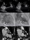
A 77-year-old female with past medical history of diabetes, dyslipidemia and atrial fibrillation presented to our service complaining of shortness of breath. She was previously treated with metoprolol, rivaroxaban and furosemide. Physical exam showed no jugular venous distention, heart auscultation highlighted a holosystolic murmur at the mitral area radiating to the axillary region, presented lower extremity edema, rest was normal. Transthoracic echocardiogram showed no contractility defects, basal hypertrophy was noted with a myocardial crypt at the inferior basal region (Fig. 1A,B, white arrow; video 1 of the supplementary data); left ventricular ejection fraction was preserved with both severe mitral and tricuspid regurgitation. Coronary angiogram did not reveal coronary artery disease, and left ventriculography showed a “2-finger shaped” image at the inferior basal region with contrast penetration between them (Fig. 1C,D; video 2 of the supplementary data). Cardiac magnetic resonance confirmed the diagnosis of myocardial cleft (Fig. 1E,F, black arrow), commonly seen as a rare congenital anomaly in patients with hypertrophic cardiomyopathy. The heart team decided to perform a bioprosthetic mitral valve replacement with tricuspid annuloplasty.
Myocardial clefts commonly present as rare incidental findings, often asymptomatic. Pathogenesis is not well known, although it is believed to occur secondary to altered myocardial wall differentiation during embryogenesis. Despite not being specific for any pathology, they occur more frequently in hypertrophic and hypertensive cardiomyopathies, even though their clinical significance remains unknown. This case highlights importance of multimodality approach to evaluate and characterize myocardial clefts when diagnosis can be challenging.


