Vertebral compression fracture is a frequent problem nowadays. In certain cases, it can be treated by performing a kyphoplasty. This percutaneous technique consists of inflating a balloon into the vertebral body followed by the infusion of orthopedic cement. This procedure seeks to achieve acceptable pain control.
We present the case of a 70 year-old woman who underwent kyphoplasty as a symptomatic treatment for back pain due to multiple lumbar vertebral fractures. The patient suffered from rheumatic valve disease (severe tricuspid regurgitation, moderate mitral stenosis and regurgitation). She also had a normal left ventricular systolic function but right ventricle dilatation with mild systolic dysfunction and estimated moderate pulmonary hypertension. An elective mitral percutaneous valvuloplasty procedure had been performed 5 years earlier leading to clinical improvement. In the follow-up, the patient presented with progressive tricuspid valve regurgitation, pulmonary hypertension and dilatation of the right cardiac cavities.
Kyphoplasty was performed in an elective manner, presenting alterations in language emission and claudication of the upper right limb in the immediate postoperative period. The symptoms resolved completely in less than 30min without the need for specific treatment.
Urgent cerebral computed tomography was performed, and cement was found in the trifurcation of the left middle cerebral artery (Fig. 1), with distal perfusion not compromised. This finding reflected a paradoxical arterial embolism.

Cement was also observed in the right lung on the chest radiograph (Fig. 1 of the supplementary data), which reflected concomitant pulmonary cement embolism. Despite this, the patient remained clinically stable without changes in the gas exchange or in her hemodynamic state even though little augmentation of estimated pulmonary systolic pressure was observed (from 55mmHg to 66mmHg) in the transthoracic echocardiogram. Dimensions of right cardiac cavities remained unchanged (right atrium of 40cm2 and right ventricle basal telediastolic diameter of 51mm). In this transthoracic echocardiogram, performed after kyphoplasty, a small atrial septal defect was observed (video 1 of the supplementary data) and a patent foramen ovale (PFO).
Oral anticoagulation was started at the discretion of the neurology team as secondary prevention for paradoxical embolisms. In addition, to support this decision, it was argued that the patient had a high risk of developing atrial fibrillation with the consequent indication of chronic oral anticoagulation. Patient was discharged with programmed follow-up in neurology outpatient consultations.
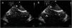
Thirty days after the orthopedic procedure, the patient went to the emergency department after presenting 2 transient cerebral ischemic episodes. The neuroimaging tests were repeated, and they did not show any changes with respect to those performed a month earlier. In a follow-up transoesophageal echocardiogram performed by an experienced echocardiographist the atrial septal defect could not be seen (video 2 of the supplementary data), but a PFO with a right-to-left shunt, a prominent Eustachian valve and an atrial septal aneurysm were observed (Fig. 2; video 3 of the supplementary data). She was discharged under the same anticoagulation regimen, but an outpatient cardiac evaluation was planned. In that consultation, the percutaneous closure of PFO was indicated as secondary prevention of paradoxical embolisms. At the time the transesophageal echocardiogram was performed, oral consent was obtained from the patient for publication of the case.

Transoesophageal echocardiogram. Agitated saline serum test with visualization of microbubbles in the right atrium that pass to the left atrium through a patent foramen ovale. Atrial septal aneurysm (asterisk). Prominent Eustachian valve (arrow). A) Ventricular systole; B) Ventricular diastole.
While waiting for the PFO closure procedure, the patient returned to emergency department due to deterioration of general condition, dizziness and vomiting. Intracerebellar hemorrhage was demonstrated in a neuroimaging study. She presented rapid clinical deterioration and died.
Cement embolisms from orthopedic surgeries are frequently seen in local veins and have also been identified in pulmonary arteries. In fact, cases of lethal pulmonary embolisms have been described.1 Besides, cases of cardiac tamponade due to perforation of right cardiac cavities by means of orthopedic cement have been described.2 Finally, cases of cerebral infarction resulting from paradoxical embolism of cement has also been reported.3 In this case we describe a stroke due to cement embolism in a patient who previously underwent mitral valvuloplasty through atrial septal puncture.
Kyphoplasty presents a risk of pulmonary embolism that ranges from 2.1% to 26%,4 so the risk of paradoxical embolism is not negligible if the patient presents a right-to-left intracardiac shunt. Then, it may be reasonable to perform a transthoracic echocardiogram with bubble test in all patients who undergo kyphoplasty to rule out intracardiac shunts.
Given the increased risk of paradoxical embolism in patients with atrial septal defects, it seems prudent to close the atrial septal defect first and perform the orthopedic procedure with cement afterwards.
Closing a PFO prior to kyphoplasty is a more controversial decision to take. Percutaneous closure of PFO is a very widespread procedure, of progressively greater use and with a low complication rate.5,6 At present there is extensive scientific evidence to indicate PFO percutaneous closure in cases of cryptogenic strokes.6 As for primary prevention, PFO closure could be indicated in the presence of major risk factors for cryptogenic stroke, such as an atrial septal aneurysm, a prominent Eustachian valve, a prominent Chiari network or a large right-to-left spontaneous shunt.6
In any case, the formation of a multidisciplinary team that includes specialists in cardiology is convenient for final decision making.
FundingNone.
Conflicts of interestNone.