

QTc interval alterations have been described in patients receiving sofosbuvir. Our objective was to evaluate the effect of anti-hepatitis C virus drugs on the human cardiac conduction system.
MethodsWe included all patients with hepatitis C virus infection that started treatment with direct-acting antivirals in a tertiary hospital between May 2016 and March 2017. Three electrocardiograms were performed in them: before starting treatment; during treatment; and at least 3 weeks after finished treatment. Heart rate, PR, QRS, and QTc intervals were compared between patients with and without sofosbuvir. Patients were followed for a mean time of 41.9 weeks.
ResultsA total of 101 patients were studied, 61 received treatment with sofosbuvir and 40 without sofosbuvir. No differences were found between the 2 groups regarding heart rate, and PR intervals. There was a statistically significant enlargement of QTc in patients with sofosbuvir at the fourth week of treatment (415.3ms vs 420.8ms) that returned to baseline values once finalized (411.1ms; P=.029). These differences were not observed in patients without sofosbuvir.
ConclusionsWe observed a statistically significant prolongation of the QTc interval at the fourth week of treatment in patients with sofosbuvir, which returned to baseline levels once the treatment was finished. Further prospective studies are needed to assess the clinical relevance of these findings.
Se han descrito alteraciones del intervalo QTc en pacientes en tratamiento con sofosbuvir. Nuestro objetivo fue evaluar el efecto de los fármacos contra el virus de la hepatitis C sobre el sistema de conducción cardiaca humana.
MétodosIncluimos a todos los pacientes con infección por VHC que iniciaron tratamiento con antivirales de acción directa en un hospital terciario entre mayo de 2016 y marzo de 2017. Se les realizaron 3 electrocardiogramas: antes de iniciar el tratamiento, durante el tratamiento y al menos 3semanas después de finalizar el tratamiento. Se compararon la frecuencia cardiaca y los intervalos PR, QRS y QTc entre pacientes con y sin sofosbuvir. Se siguió a los pacientes durante un tiempo medio de 41,9semanas.
ResultadosSe estudió a 101 pacientes, de los que 61 recibieron tratamiento con sofosbuvir y 40 sin sofosbuvir. No se encontraron diferencias entre los 2 grupos en cuanto a la frecuencia cardiaca y los intervalos PR. Se observó un aumento estadísticamente significativo del intervalo QTc en los pacientes con sofosbuvir en la cuarta semana de tratamiento (415,3ms frente a 420,8ms), que volvió a los valores basales una vez finalizado el tratamiento (411,1ms; p=0,029). Estas diferencias no se observaron en los pacientes sin sofosbuvir.
ConclusionesObservamos una prolongación estadísticamente significativa del intervalo QTc en la cuarta semana de tratamiento en los pacientes con sofosbuvir, que volvió a los niveles basales una vez finalizado el tratamiento. Se necesitan más estudios prospectivos para evaluar la relevancia clínica de estos hallazgos.
Direct-acting antivirals (DAA) against hepatitis C virus (HCV) have changed the paradigm of HCV infection, showing a high efficacy in treatment with a sustained viral response rate greater than 95%.1 Different families of drugs make up the framework of the new antivirals: the inhibitors of NS3 (protease), NS5A and NS5B (polymerases). Although the standards of care for HCV are continuously changing, sofosbuvir keeps being the backbone of the treatment.2,3 The rate of side effects of this new DAA is less than 10%. The most frequent are headache, fatigue, nausea, and diarrhea.4
However, in January 2015, the French National Agency for Medicines and Health Products Safety declared severe symptomatic events of bradyarrhythmia and syncope in patients treated with sofosbuvir and daclatasvir in combination with amiodarone.5 In March 2015, the Food and Drug Administration reported 9 patients under treatment with amiodarone combined with sofosbuvir and other DAA (ledipasvir and daclatasvir) who developed symptomatic bradyarrhythmias.6 One of them died and 3 required implantations of a pacemaker. In the remaining patients, the suspension of antiviral treatment was accompanied by symptomatic resolution, and in 3 of them the reintroduction of sofosbuvir induced the recurrence of symptomatic bradycardia.
Additionally, new cases of sinus node dysfunction, atrio-ventricular blockade, or non-sustained ventricular tachycardia (NSVT) have been reported in patients under combinations of sofosbuvir and other DAA but without association with amiodarone.7
Basic research studies in animal models have unsuccessfully tried to elucidate the underlying pathophysiological mechanism of these concomitant effects,8–12 and the effects of sofosbuvir and other DAA on the human cardiac conduction system remains also unknown. A study including 39 patients has suggested that there may be a lengthening of the QT interval in patients treated with sofosbuvir without amiodarone, specifically during the first week of treatment.13 Another study suggested prolongation of QTc during treatment with a combination of sofosbuvir and ledipasvir in 395 patients with liver fibrosis.14
Thus, our study aims to describe the effect of the new antivirals on the cardiac conduction system; to check if these changes persist once the treatment is completed; to assess the safety of their administration in combination with drugs that act directly on the cardiac conduction system, and to determine possible adverse clinical events during treatment.
MethodsThe study protocol conformed to the ethical guidelines of the 1975 Declaration of Helsinki and was approved by the Ethics Committee of Hospital Clínico San Carlos, Madrid. Informed consent in writing was obtained from each patient. This prospective observational cohort study was carried out entirely in a tertiary care referral center.
Study populationWe consecutively included all patients with diagnosis of HVC infection between May 2016 and March 2017 who were going to start treatment with DAA. Whether sofosbuvir was used as part of the combination of drugs for the treatment or not was up to the decision of the treating physician, according to the EASL clinical practice guidelines in force during recruitment.15 Patients were excluded from the study if they were under 18 years of age or withdrew consent.
We collected the following baseline variables: demographic variables related to the epidemiology of the infection (genotype of the HCV, grade of liver by hepatic elastography, viral load, presence of cryoglobulinemia, MELD score), cardiovascular risk factors and diseases, concomitant treatment with antiarrhythmics, including betablockers, and other drugs that could potentially lengthen the QTc interval. The combination of antiviral drugs for the treatment of virus C was also collected.
ElectrocardiogramsAll patients underwent 3 electrocardiograms (ECG): the first one was performed before starting the antiviral treatment (basal); the second, during treatment, 4 weeks after the start; and the third, at least 3 weeks after it was finalized, considering that the duration of treatment could be of 8, 12 or 24 weeks.
PageWriter TC30 Cardiograph (Philips, United States) device was used to obtain the ECG. Twelve lead electrocardiograms were obtained in supine at a speed of 25mm/s and a gain of 10mm/mV.
The following electrocardiographic variables were collected: type of rhythm, heart rate, duration of the PR interval (normal <200ms), duration of the QRS complex (normal <120ms), duration of the QT interval and corrected QT interval (QTc), normal <450ms in men and <470ms in women. All measurements were made manually on paper by an observer blinded to the patient's clinical data.
For the measurement of the ECG intervals, the mean of the longest measurement in precordial leads (preferably in lead II) and limb leads (preferably in lead V3) was obtained. The QT interval was defined as the distance from the beginning of the QRS to the intersection of a tangent drawn to the steepest last limb of the T-wave with the baseline as defined by Lepeschkin and Surawicz.16 The Bazet method was used to calculate the QTc interval given that it is the most widely used method to correct the QT interval, and that extreme heart rates, where its performance is worse, were not anticipated.
Follow-upPatients were followed up by telephone calls and by searching in their medical history to determine admissions to the Emergency Department or consultations to their general practitioner for palpitations, dizziness, syncope, chest pain or other reasons.
Statistical analysisThe normal distribution of the variables was checked using the Shapiro–Wilk test for normality. Continuous variables are represented as mean and standard deviation (SD) or median and interquartile range. Dichotomous variables were compared with a 2-tailed t-test, or Fisher–Pitman permutation test when necessary, and in cases with more than two groups the analysis of variance (ANOVA), or multivariate analysis of variance (MANOVA) test were used. Categorical variables are expressed as number and percentage and were compared with a Chi squared test. For the analysis of quantitative variables in subgroups the nonparametric randomization test of Fisher–Pitman was used.
All tests were 2-sided, and differences were considered statistically significant at P-values <.05. Statistical analysis was performed with Stata V.12.0 (StataCorp, United States).
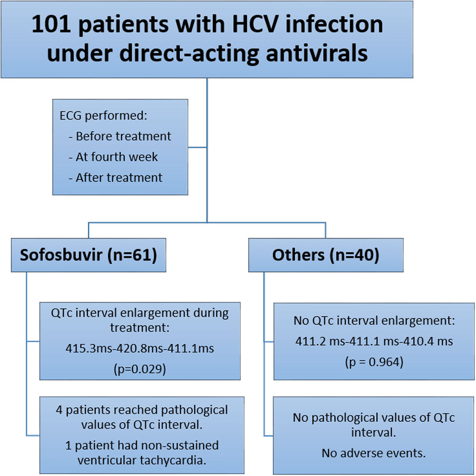
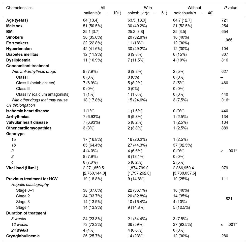
ResultsPatient characteristicsWe included 115 patients in the study, of which 101 had a complete follow-up with the 3 ECG performed (Fig. 1). In 14 patients all the 3 ECG were not achieved for the following reasons: 2 patients died during follow-up; 2 moved to another region; in 2 patients follow-up was lost, and 7 did not attend the appointments to perform any of the ECG for other reasons. The baseline characteristics of patients are shown in Table 1.
Basal characteristics of all patients and patients with and without sofosbuvir.
| Characteristics | All patients(n=101) | With sofosbuvir(n=61) | Without sofosbuvir(n=40) | P value |
|---|---|---|---|---|
| Age (years) | 64 [13.4] | 63.5 [13.9] | 64.7 [12.7] | .721 |
| Male sex | 51 (50.5%) | 30 (49.2%) | 21 (52.5%) | .254 |
| BMI | 25.1 [3.7] | 25.2 [3.8] | 25 [3.5] | .654 |
| Smokers | 36 (35.6%) | 20 (32.8%) | 16 (40%) | .066 |
| Ex smokers | 22 (22.8%) | 11 (18%) | 12 (30%) | |
| Hypertension | 42 (41.6%) | 30 (49.2%) | 12 (30%) | .104 |
| Diabetes mellitus | 12 (11.9%) | 6 (9.8%) | 6 (15%) | .807 |
| Dyslipidemia | 11 (10.9%) | 7 (11.5%) | 4 (10%) | .816 |
| Concomitant treatment | ||||
| With antiarrhythmic drugs | 8 (7.9%) | 6 (9.8%) | 2 (5%) | .627 |
| Class I | 0 (0%) | 0 (0%) | 0 (0%) | – |
| Class II (betablockers) | 7 (6.9%) | 5 (8.2%) | 2 (5%) | .460 |
| Class III | 0 (0%) | 0 (0%) | 0 (0%) | – |
| Class IV (calcium antagonists) | 1 (1%) | 1 (1.6%) | 0 (0%) | .440 |
| With other drugs that may cause QT prolongation | 18 (17.8%) | 15 (24.6%) | 3 (7.5%) | .016* |
| Ischemic heart disease | 1 (1%) | 1 (1.6%) | 0 (0%) | .440 |
| Arrhythmias | 7 (6.93%) | 6 (9.8%) | 1 (2.5%) | .134 |
| Valvular heart disease | 7 (6.93%) | 5 (8.2%) | 1 (2.5%) | .134 |
| Other cardiomyopathies | 3 (3%) | 2 (3.3%) | 1 (2.5%) | .889 |
| Genotype | ||||
| 1a | 17 (16.8%) | 16 (26.2%) | 1 (2.5%) | <.001* |
| 1b | 65 (64.4%) | 27 (44.3%) | 37 (92.5%) | |
| 2 | 4 (4.0%) | 4 (6.6%) | 0 (0%) | |
| 3 | 8 (7.9%) | 8 (13.1%) | 0 (0%) | |
| 4 | 8 (7.9%) | 5 (8.2%) | 2 (5%) | |
| Viral load (UI/mL) | 2.271,659.5 [2,769,144.0] | 1,874,799.0 [1,797,262.0] | 2,866,950.4 [3,738,037.6] | .079 |
| Previous treatment for HCV | 19 (18.8%) | 9 (14.8%) | 10 (25%) | .111 |
| Hepatic elastography | ||||
| Stage 0–1 | 38 (37.6%) | 22 (36.1%) | 16 (40%) | .821 |
| Stage 2 | 34 (33.7%) | 20 (32.8%) | 14 (35%) | |
| Stage 3 | 14 (13.9%) | 10 (16.4%) | 4 (10%) | |
| Stage 4 | 14 (13.9%) | 9 (14.8%) | 5 (12.5%) | |
| Duration of treatment | ||||
| 8 weeks | 24 (23.8%) | 21 (34.4%) | 3 (7.5%) | <.001* |
| 12 weeks | 73 (72.3%) | 36 (59%) | 37 (92.5%) | |
| 24 weeks | 4 (4%) | 4 (6.6%) | 0 (0%) | |
| Cryoglobulinemia | 26 (25.7%) | 14 (23%) | 12 (30%) | .280 |
Values are n (%) or mean [SD]. BMI, body mass index; HVC, hepatitis C virus; MELD, model end-stage liver disease.
Concomitant cardiovascular diseases were collected: only 1 patient (1%) had ischemic heart disease, 6.9% arrhythmias (all of them were supraventricular), 6% had some degree of valve heart disease, and 3 patients had a history of other heart diseases (1 patient had tako-tsubo syndrome, another dissecting aneurysm of ascending aorta treated with an ascending aortic graft, and another had had acute pericarditis).
According to the Vaughan–Williams classification of antiarrhythmic drugs, only 1 patient was on non-dihydropyridine calcium channel blockers (class IV), 6.95% took betablockers (class II), and no patients were on class I or III antiarrhythmic drugs.
Grade of liver fibrosis by hepatic elastography before starting treatment was determined as F0–F1 in 37.6% of patients, F2 in 33.7% of patients, F3 in 13.9%, and F4 in 13.9%. Before starting treatment, 81.1% had genotype 1 (16.8% had genotype 1a and 64.4% genotype 1b), 4% had genotype 2, 7.9% had genotype 3, and 7.9% had genotype 4. One patient had double genotype (1b and 4). Median MELD score was 7.24±3. Median viral load was 2,271,659.5±2,769,144.1UI/mL. Cryoglobulinemia was present in 25.7% of patients, and 18.8% had been previously treated against HVC. Treatment duration was 8 weeks in 23.8% of patients, 12 weeks in 72.3% and 24 weeks in 4%. At the time of the third ECG, 95% of patients had sustained viral response.
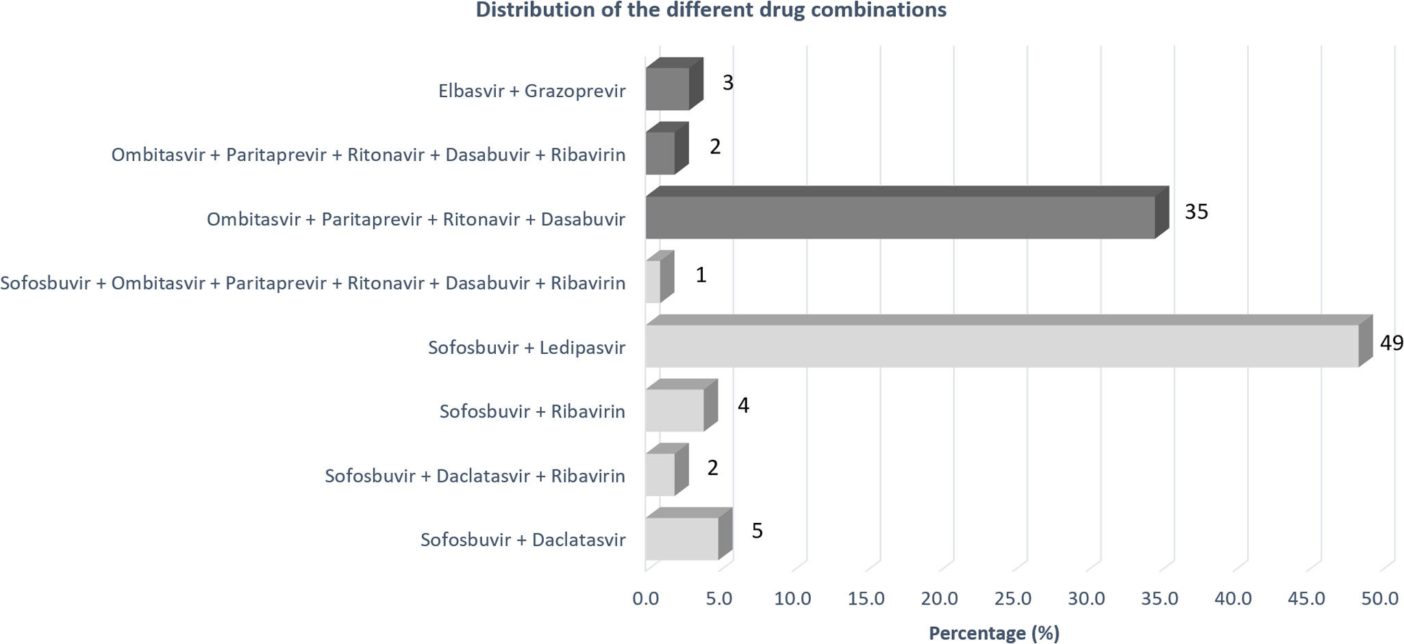
Anti-hepatitis C virus treatmentOf the 101 patients who completed the study, 60.4% received treatment with sofosbuvir in any of their combinations (sofosbuvir+ledipasvir 48.5%, sofosbuvir+daclatasvir 6.9%, sofosbuvir without another DAA 4%, sofosbuvir+ombitasvir, paritaprevir, ritonavir, dasabuvir 1%); 11.6% of patients treated with sofosbuvir also received ribavirin (n=7).
On the other hand, 36.6% received the combination of ombitasvir, paritaprevir, ritonavir, dasabuvir (5.4% of these patients also received ribavirin) and 3% received treatment with elbasvir plus grazoprevir (none of them associated with ribavirin). The pharmacological combinations are summarized in Fig. 2.
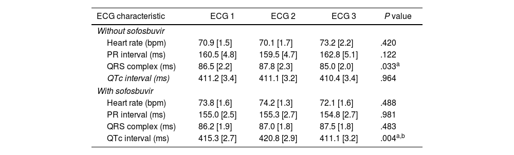
Electrocardiogram findingsIn the basal ECG, 1 patient had atrial flutter with pacemaker-mediated ventricular stimulation and 2 had atrial fibrillation. The remaining patients were in sinus rhythm. One of them had frequent supraventricular extrasystole. The second ECG was performed 29.6 days on average after the start of treatment (standard deviation [SD], 8), and the third ECG was performed 111.4 days after the end of treatment (SD, 80.3). There were 2 patients who had sinus rhythm in the first ECG and presented atrial fibrillation in the second ECG. Mean heart rate, PR interval, QRS complex and QTc interval for each ECG are presented in Table 2.
Comparison of the measurements in the 3 ECG in groups without and with sofosbuvir.
| ECG characteristic | ECG 1 | ECG 2 | ECG 3 | P value |
|---|---|---|---|---|
| Without sofosbuvir | ||||
| Heart rate (bpm) | 70.9 [1.5] | 70.1 [1.7] | 73.2 [2.2] | .420 |
| PR interval (ms) | 160.5 [4.8] | 159.5 [4.7] | 162.8 [5.1] | .122 |
| QRS complex (ms) | 86.5 [2.2] | 87.8 [2.3] | 85.0 [2.0] | .033a |
| QTc interval (ms) | 411.2 [3.4] | 411.1 [3.2] | 410.4 [3.4] | .964 |
| With sofosbuvir | ||||
| Heart rate (bpm) | 73.8 [1.6] | 74.2 [1.3] | 72.1 [1.6] | .488 |
| PR interval (ms) | 155.0 [2.5] | 155.3 [2.7] | 154.8 [2.7] | .981 |
| QRS complex (ms) | 86.2 [1.9] | 87.0 [1.8] | 87.5 [1.8] | .483 |
| QTc interval (ms) | 415.3 [2.7] | 420.8 [2.9] | 411.1 [3.2] | .004a,b |
Values are mean [SD]. ECG, electrocardiogram; bpm, beats per minute; ms, miliseconds; QTc, corrected QT interval.
There were no significant differences in heart rate, or PR interval between the first, the second and the third ECG, neither in the whole group, nor in patients under treatment with sofosbuvir or other drugs combinations. In the group of patients without sofosbuvir, we found a small but statistically significant difference in the QRS complex between the second and the third ECG (86.5ms vs 87.8ms vs 85.0ms; P=.033).
Conversely, the QTc interval was significantly prolonged between the first and the second ECG in the group under sofosbuvir, returning to basal values in the third ECG after finishing the treatment (415.3ms vs 420.8ms vs 411.2ms; P=.029). On the contrary, in the group without sofosbuvir, no significant differences were found in QTc duration between the 3 ECG (411.2ms vs 411.1ms vs 410.4ms; P=.964).
Pathological QTc intervalOnly 1 female patient had a pathological QTc interval before treatment, which remained pathological during and after treatment. This patient received treatment without sofosbuvir.
In addition, there were 4 male patients in the sofosbuvir group who, having normal QTc values in the basal ECG, reached QTc pathological values during the treatment. In these 4 patients, the QTc interval in the third ECG returned to normal values. In the group without sofosbuvir there were no patients with pathological QTc lengthening during the treatment.
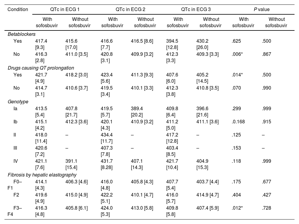
Subgroup analysis of QTcTo further evaluate other potential factors that could affect the QTc interval, we compared the variations of the QTc interval in different subgroups (concomitant use of betablockers, drugs causing QTc prolongation, genotype and grade of liver fibrosis by hepatic elastography). All subgroups showed a tendency of enlarging the QTc in patients under sofosbuvir treatment, despite the difference remained only significant in patients without betablockers, with drugs causing QTc prolongation, and patients with grade F3–F4 of fibrosis. In patients without sofosbuvir there were no differences in any subgroup. Subgroup analysis is shown in Table 3.
QTc subgroup analysis. Variation of QTc depending on concomitant treatment, genotype and grade of liver fibrosis.
| Condition | QTc in ECG 1 | QTc in ECG 2 | QTc in ECG 3 | P value | ||||
|---|---|---|---|---|---|---|---|---|
| With sofosbuvir | Without sofosbuvir | With sofosbuvir | Without sofosbuvir | With sofosbuvir | Without sofosbuvir | With sofosbuvir | Without sofosbuvir | |
| Betablockers | ||||||||
| Yes | 417.4 [9.3] | 415.6 [17.0] | 416.6 [7.7] | 416.5 [8.6] | 394.5 [12.8] | 430.2 [26.0] | .625 | .500 |
| No | 416.3 [2.8] | 411.0 [3.5] | 420.8 [3.1] | 409.9 [3.2] | 412.3 [3.3] | 409.3 [3.3] | .006* | .867 |
| Drugs causing QT prolongation | ||||||||
| Yes | 421.7 [4.9] | 418.2 [3.0] | 423.4 [5.6] | 411.3 [9.3] | 407.6 [6.0] | 405.2 [14.5] | .014* | .500 |
| No | 414.7 [3.1] | 410.6 [3.7] | 419.5 [3.4] | 410.1 [3.3] | 412.3 [3.8] | 410.8 [3.5] | .070 | .990 |
| Genotype | ||||||||
| Ia | 413.5 [5.4] | 407.8 [21.7] | 419.5 [5.7] | 389.4 [20.2] | 409.8 [6.4] | 396.6 [21.6] | .299 | .999 |
| Ib | 415.1 [4.2] | 412.3 [3.6] | 420.1 [4.3] | 410.9 [3.2] | 411.2 [5.0] | 411.1 [3.6] | .0.168 | .915 |
| II | 418.0 [11.4] | – | 434.4 [11.7] | – | 417.2 [12.8] | – | .125 | – |
| III | 420.6 [7.2] | – | 407.3 [7.8] | – | 403.4 [8.5] | – | .153 | – |
| IV | 421.1 [7.6] | 391.1 [15.4] | 431.7 [8.28] | 407.1 [14.3] | 421.7 [10.4] | 404.9 [15.3] | .118 | .999 |
| Fibrosis by hepatic elastography | ||||||||
| F0–F1 | 414.1 [4.3] | 406.3 [4.6] | 416.0 [4.8] | 405.8 [4.3] | 407.7 [5.4] | 403.7 [4.4] | .175 | .677 |
| F2 | 419.6 [4.9] | 415.0 [4.9] | 422.2 [5.1] | 410.1 [4.7] | 416.0 [5.7] | 414.9 [4.7] | .404 | .427 |
| F3–F4 | 416.3 [4.8] | 405.8 [6.1] | 424.0 [5.3] | 413.0 [5.8] | 409.8 [5.8] | 407.4 [5.9] | .012* | .728 |
Values are mean [SD]. ECG, electrocardiogram; QTc, corrected QT interval.
The mean time of follow-up was 41.9±14 weeks. During treatment, in the group with sofosbuvir, 1 patient consulted the emergency room or his general practitioner due to palpitations, 1 due to dizziness and 1 due to chest pain. This last one was referred to the cardiology outpatient clinic. A Holter ECG was performed during HCV treatment where frequent ventricular premature ventricular beats and NSVT were observed. After finishing HCV treatment, and up-titrating betablockers, premature ventricular beats rate remained similar. Three patients also presented occasional palpitations but did not consult for it.
In the group without sofosbuvir, 1 patient consulted for dizziness and 1 due to chest pain. No studies were performed. In this group there were also 2 patients with occasional palpitations who did not consult. No patient presented syncope during the follow-up. The cause of death of the 2 patients who died during the follow-up was oncological. Both of them died several months after HCV treatment was finished. One of them had a large B-cell lymphoma that invaded the right atrium and caused complete atrio-ventricular block. The second had a glioblastoma multiforme.
DiscussionIn our study, we have observed a statistically significant lengthening of the QTc interval at the fourth week of treatment in patients undergoing treatment with sofosbuvir that returned to baseline values in the ECG performed once the treatment was finished. This lengthening did not happen in patients taking other DAA drugs.
Regarding the association between DAA and cardiac conduction system abnormalities, in 2015 several cases of symptomatic bradyarrhythmia with DAA in combination with amiodarone were reported,5,6 and since then, this combination is discouraged. More recently, new cases of symptomatic sinus node dysfunction, atrio-ventricular blockade, and NSVT have been described in patients under combination of sofosbuvir and other DAA but without amiodarone.7 In vivo experimental studies have demonstrated the appearance of bradycardia and arterial hypotension in rhesus monkeys and guinea pigs when co-administering sofosbuvir and amiodarone, although these effects did not occur when the drugs were administered separately.8 An in vitro model has shown that the combination amiodarone-sofosbuvir interacts with intracellular calcium-handling mechanisms, similar to the action of calcium channel blockers.10 However, no structural or electrophysiological disturbance has been described in humans taking sofosbuvir without amiodarone.
Biomy et al.17 studied 170 patients who were treated with sofosbuvir and underwent a cardiological follow-up with electrocardiogram and echocardiogram to evaluate the potential cardiovascular effects of DAA. Three ECG were performed, at baseline and at 6- and 12-month follow-up, and no significant changes were observed in the QTc interval or other electrocardiographic parameters, although they did not perform any ECG during the treatment.
Durante-Mangoni et al.13 carried out a study in 39 patients with DAA, of whom 26 received sofosbuvir, potential alterations of the cardiac conduction system were analyzed. The authors performed a basal ECG, and weekly additional ECG during the first month of treatment. Consistent with our findings, they did not find any significant differences in heart rate, PR interval or QRS complex duration. Interestingly, they found a significant lengthening of the QTc interval during the first week of treatment with sofosbuvir, which disappeared in the fourth week of treatment.
In contrast to this last finding, our results showed an elongation of the QTc interval at the fourth week of treatment in patients with sofosbuvir that was not found in patients without sofosbuvir and corrected to baseline values after finishing the treatment. This disparity may be due to a larger number of patients in our study but since the timing of the ECG performance is different in both studies, we cannot come to a conclusion about this discordance.
Tahata et al.14 found newly developed QTc prolongation in 6.4% in a large cohort of Japanese patients during treatment with sofosbuvir/ledipasvir, returning to baseline at week 12 after treatment in consonance with our study, but did not study this effect with other therapeutic groups.
When we analyzed different subgroups that could potentially influence these electrical alterations, we found a trend towards lengthening of the QT interval in all subgroups taking sofosbuvir. In patients who took sofosbuvir but either did not take betablockers or took drugs causing QT prolongation, it should be noted that the most notable differences were found between the second ECG and the third ECG, with the third QTc being even shorter than the baseline one. However, the small number of patients per group limits the value of these findings. In patients without sofosbuvir, there were not differences in any of the subgroups.
Treatment duration is different in both groups. One third of patients in the sofosbuvir group received a shortened 8-week regimen, compared with only 3 patients (7.5%) in the non-sofosbuvir group. However, the baseline ECG was performed before starting any treatment and the ECG during treatment was always performed with the same treatment duration (4 weeks) in all cases, so treatment duration is not expected to be a factor influencing the electrocardiographic differences acquired during treatment compared to baseline.
Regarding the clinical impact of the rise in QTc duration in patients taking sofosbuvir, in our study there were 4 patients (4%) who reached pathological values of QTc interval duration during the treatment and returned to non-pathological values once the treatment with sofosbuvir was finished. None of them had any clinical event during treatment.
In addition, during follow-up, in 1 of the patients in the sofosbuvir group, NSVT and multiple ventricular extrasystole were documented. In the same way, Nirei et al.7 recently published 2 cases of patients under treatment with sofosbuvir who presented NSVT. In the first case, the NSVT occurred 23min after the first dose of sofosbuvir/ledipasvir. In the second case, the NSVT occurred 2 days after the start of the treatment and was repeated 5 days later. In both cases, the drug was subsequently withdrawn and a cardiological study was carried out prior to hospital discharge, without revealing any pathological finding. Unfortunately, our patient was not further studied.
On the other hand, in the studies of Biomy et al.17 and Durante-Mangoni et al.,13 no significant clinical cardiac event during the follow-up of patients was registered.
Most of the drugs that lengthen the QT interval (among them, amiodarone) are well known. These drugs confer a potential risk of generation of polymorphic ventricular tachycardia which, if sustained, is highly fatal. Therefore, certain precautions must be taken when using these drugs, such as avoiding concomitant use of 2 drugs that can prolong QT interval, avoiding the administration in patients with congenital long QT syndrome, monitoring QT in some cases, and sometimes even withdraw these drugs.
Therefore, this study raises the possible recommendation of performing electrocardiographic monitoring at baseline and during the first few weeks of treatment (for example in week 4) as well as trying to avoid treatment with concomitant drugs that can lengthen the QTc interval.
LimitationsThis study has several limitations. First, our findings are based on punctual ECG measurements at certain times of treatment and after treatment, which did not allow us to determine the precise moment in which the QTc interval starts to prolong, and when these changes disappear. In addition, the absence of continuous monitoring prevents diagnosing the presence of non-sustained arrhythmias.
Secondly, due to the limited sample size, no definite conclusions can be drawn about whether the effect of QT elongation is due to any particular combination of drugs including sofosbuvir, nor if there are additive effects with other drugs that can potentially lengthen the QTc. Furthermore, there are new drug combinations currently available that were not marketed at the time of recruitment in this study, so we do not know the electrocardiographic effects of these combinations.
Finally, since it is an observational study, it is not possible to establish causal relationships between treatment with sofosbuvir and the rise in QTc duration, although the fact that the QTc returns to normal values after treatment fulfils another of the main pharmacological criteria of causality (reversibility). Thus, a prospective study with continuous monitoring in patients taking sofosbuvir would help to determine the appearance of QTc lengthening and the presence of adverse non-sustained arrhythmic events.
ConclusionsPatients on treatment with sofosbuvir, a statistically significant prolongation of the QTc interval was observed at the fourth week of treatment, which returned to baseline values once the treatment was finished. In 6.6% of patients with sofosbuvir, the QTc interval reached pathological values. In those patients under treatment with other DAA, no significant changes in QTc duration were found. Further prospective studies are needed to confirm and evaluate the clinical relevance of these findings.
- –
DAA against HCV have changed the paradigm of HCV infection, showing a high efficacy.
- –
QTc interval alterations have been described in patients receiving sofosbuvir.
- –
We observed a statistically significant prolongation of the QTc interval at the fourth week of treatment in patients with sofosbuvir, which returned to baseline levels once the treatment was finished.
- –
These alterations did not happen with other therapeutic groups.
- –
With these findings we suggest avoiding the combination of sofosbuvir with drugs that can prolong the QT interval.
None declared.
Ethical considerationsThe study protocol was in accordance with the ethical standards of the 1975 Declaration of Helsinki and was approved by the Ethics Committee of Hospital Clínico San Carlos, Madrid. Written informed consent was obtained from each patient. In accordance with SAGER guidelines, we confirm that potential sex and gender biases have been taken into account in the preparation of this article.
Declaration on the use of artificial intelligenceNo artificial intelligence was used in the design, analysis, preparation or writing of this manuscript.
Authors’ contributionsO. Neva López-García and D. García-Arribas contributed to make the conception, the design of the study, the acquisition of data, and drafting the article. C. Olmos has contributed to making the design of the study and the statistical analysis and interpretation of data. M.J. Devesa Medina has contributed to making the design of the study and revising it critically for important intellectual content. J. Higueras Nafría has contributed to reviewing it critically for important intellectual content. S. Izquierdo Rubio, F. Cuenca Alarcon, M. Maroto Castellanos, A. Fernández-Vega, A. Cruz-Utrilla, P. Martínez-Vives, and E. Rey contributed to the final approval of the version to be submitted. All authors read and approved the final version of the paper.
Conflict of interestNone of the authors have any conflicts of interest.