A 38-year-old female patient was referred to the emergency department due to chest pain and an ambulatory transthoracic echocardiogram describing an important mobile mass in the right atrium (RA).
The differential diagnosis included a thrombus, a right atrial myxoma/other primary cardiac tumor or a tumor infiltrating the vena cava from a nearby organ.
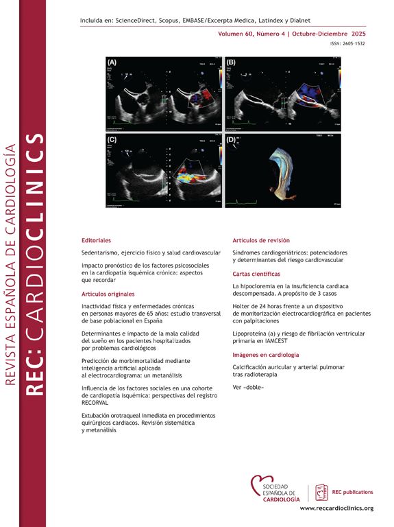
The transesophageal echocardiogram showed a worm-like, very elongated, filiform mass of significant dimensions that seemed to emerge from the inferior vena cava, without any attachment to other cardiac structures, occupying the RA and prolapsing to the right ventricle during diastole (Fig. 1A–C, white arrow).
Cardiovascular magnetic resonance (CMR) showed a filiform mass, now clearly seen to be implanted in the inferior vena cava, near the junction with the RA, without any evidence of venous invasion from nearby structures (Fig. 1D–E). This lesion did not have any signs of vascularity in the perfusion sequences and no evidence of enhancement in early or delayed enhancement sequences (Fig. 1F–H, yellow arrow). The CMR findings were compatible with a thrombus.
Because thrombolysis of giant thrombi may be ineffective, patients in this situation may require surgery which could be the safest option.
While waiting for surgery, the patient suffered a massive pulmonary embolism. She underwent thrombolysis with good clinical response. A follow-up transthoracic echocardiogram failed to show any significant mass on the RA.
Subsequent study confirmed the presence of a prothrombotic state, with the diagnosis of antiphospholipid syndrome.
This unusual case illustrates the importance of CMR in studying the nature of cardiac masses.


