Purulent pericarditis has become an uncommon disease since the antibiotic era and improvements in hygienic conditions, accounting for less than 1% of pericarditis cases today in high-income countries.1,2
In the pre-antibiotic era, it primarily affected children and young adults; however, since the mid-20th century, it has increasingly affected adults.2 At the beginning of the 20th century, the disease was commonly associated with a contiguous infectious disease, most frequently pneumococcal pneumonia. In the latter part of the 20th century, the prevalence of staphylococcal infections, particularly Staphylococcus aureus, increased, mainly in patients with high comorbidity, spreading to the pericardium from distant sites.2,3
To our knowledge, in the 21st century, there have been only a few case series,4 leaving its etiology, presentation, and prognosis poorly understood. Our study aims to describe current underlying causes, at-risk populations, and outcomes.
This retrospective observational study included all consecutive adult patients admitted with a diagnosis of purulent pericarditis between 2010 and 2022 at a tertiary referral center (Hospital Vall d’Hebron, Barcelona, Spain).
Patients were identified from the hospital's discharge database based on the ICD codes [I30-1 (infective pericarditis), 420.99 (other acute pericarditis) and 420.9 (acute pericarditis, unspecified)].
From the resulting list, individuals were selected if they met at least two of the following criteria: (a) macroscopic purulent fluid; (b) positive cultures of pericardial or pleural effusion; (c) predominance of polymorphonuclear infiltrate (>75%) and low glucose levels (<60mg/dL) in pericardial fluid. Patients with tuberculous pericarditis were excluded.
All patients were treated by a multidisciplinary team, considering the etiology, source, and extent of the infection.
Variables were obtained using local and national electronic health records. Categorical variables are presented as counts and percentages (%) and continuous variables as median [interquartile range].
The study was conducted in accordance with the Declaration of Helsinki and was approved by the local Institutional Review Board PR(AG)556/2023.
During the study, 17 patients with purulent pericarditis were identified. After excluding 1 with tuberculosis-related pericarditis, 16 patients were analyzed.
The median age at admission was 60.4 years [52.5–75.4], with 50% of the patients being women. Thirteen patients (81%) had an underlying chronic debilitating condition, with risky alcohol consumption being the most common. The median Charlson Comorbidity Index was 3 [2.3–4.8] points. The remaining underlying conditions and baseline characteristics are described in Tables 1 and Table 2.
Characteristics of the patients, signs, symptoms and outcome.
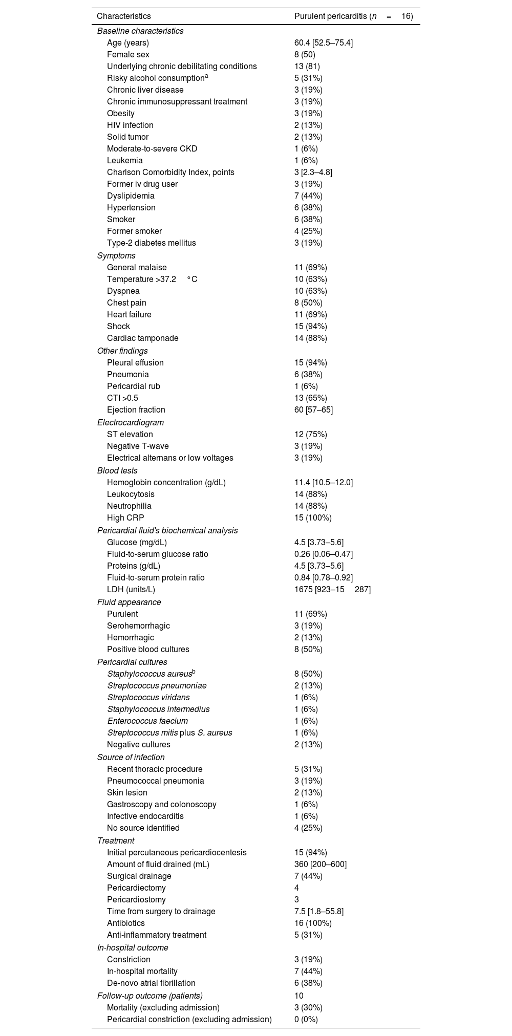
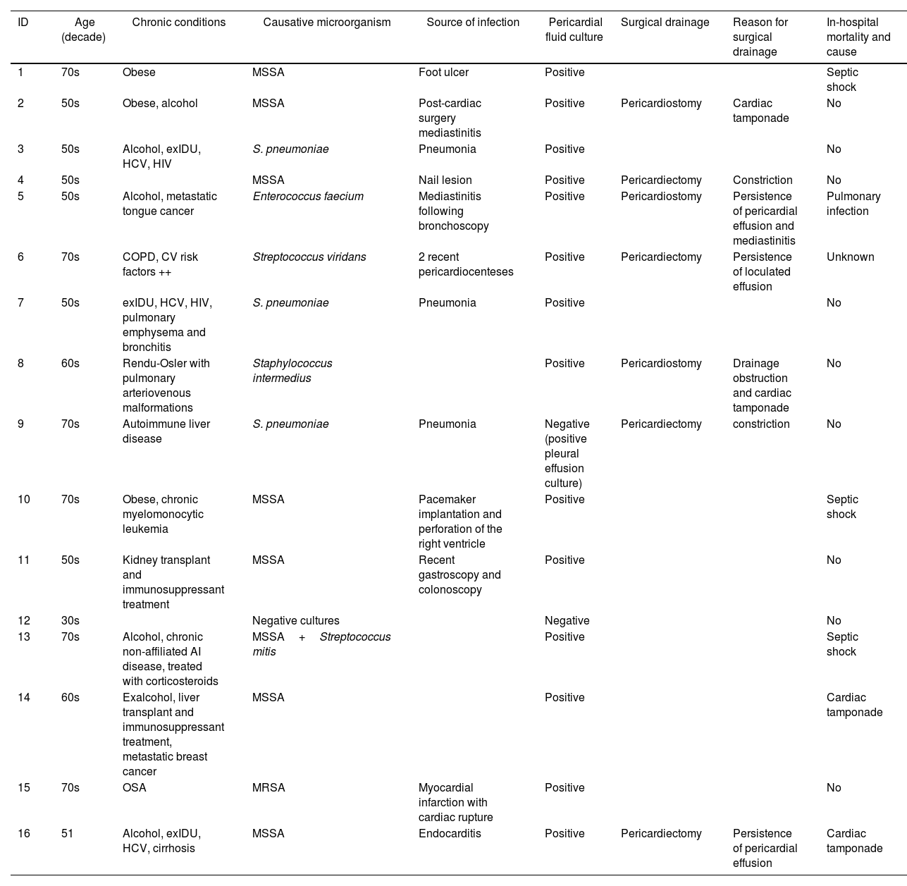
Characteristics | Purulent pericarditis (n=16) |
---|---|
Baseline characteristics | |
Age (years) | 60.4 [52.5–75.4] |
Female sex | 8 (50) |
Underlying chronic debilitating conditions | 13 (81) |
Risky alcohol consumptiona | 5 (31%) |
Chronic liver disease | 3 (19%) |
Chronic immunosuppressant treatment | 3 (19%) |
Obesity | 3 (19%) |
HIV infection | 2 (13%) |
Solid tumor | 2 (13%) |
Moderate-to-severe CKD | 1 (6%) |
Leukemia | 1 (6%) |
Charlson Comorbidity Index, points | 3 [2.3–4.8] |
Former iv drug user | 3 (19%) |
Dyslipidemia | 7 (44%) |
Hypertension | 6 (38%) |
Smoker | 6 (38%) |
Former smoker | 4 (25%) |
Type-2 diabetes mellitus | 3 (19%) |
Symptoms | |
General malaise | 11 (69%) |
Temperature >37.2°C | 10 (63%) |
Dyspnea | 10 (63%) |
Chest pain | 8 (50%) |
Heart failure | 11 (69%) |
Shock | 15 (94%) |
Cardiac tamponade | 14 (88%) |
Other findings | |
Pleural effusion | 15 (94%) |
Pneumonia | 6 (38%) |
Pericardial rub | 1 (6%) |
CTI >0.5 | 13 (65%) |
Ejection fraction | 60 [57–65] |
Electrocardiogram | |
ST elevation | 12 (75%) |
Negative T-wave | 3 (19%) |
Electrical alternans or low voltages | 3 (19%) |
Blood tests | |
Hemoglobin concentration (g/dL) | 11.4 [10.5–12.0] |
Leukocytosis | 14 (88%) |
Neutrophilia | 14 (88%) |
High CRP | 15 (100%) |
Pericardial fluid's biochemical analysis | |
Glucose (mg/dL) | 4.5 [3.73–5.6] |
Fluid-to-serum glucose ratio | 0.26 [0.06–0.47] |
Proteins (g/dL) | 4.5 [3.73–5.6] |
Fluid-to-serum protein ratio | 0.84 [0.78–0.92] |
LDH (units/L) | 1675 [923–15287] |
Fluid appearance | |
Purulent | 11 (69%) |
Serohemorrhagic | 3 (19%) |
Hemorrhagic | 2 (13%) |
Positive blood cultures | 8 (50%) |
Pericardial cultures | |
Staphylococcus aureusb | 8 (50%) |
Streptococcus pneumoniae | 2 (13%) |
Streptococcus viridans | 1 (6%) |
Staphylococcus intermedius | 1 (6%) |
Enterococcus faecium | 1 (6%) |
Streptococcus mitis plus S. aureus | 1 (6%) |
Negative cultures | 2 (13%) |
Source of infection | |
Recent thoracic procedure | 5 (31%) |
Pneumococcal pneumonia | 3 (19%) |
Skin lesion | 2 (13%) |
Gastroscopy and colonoscopy | 1 (6%) |
Infective endocarditis | 1 (6%) |
No source identified | 4 (25%) |
Treatment | |
Initial percutaneous pericardiocentesis | 15 (94%) |
Amount of fluid drained (mL) | 360 [200–600] |
Surgical drainage | 7 (44%) |
Pericardiectomy | 4 |
Pericardiostomy | 3 |
Time from surgery to drainage | 7.5 [1.8–55.8] |
Antibiotics | 16 (100%) |
Anti-inflammatory treatment | 5 (31%) |
In-hospital outcome | |
Constriction | 3 (19%) |
In-hospital mortality | 7 (44%) |
De-novo atrial fibrillation | 6 (38%) |
Follow-up outcome (patients) | 10 |
Mortality (excluding admission) | 3 (30%) |
Pericardial constriction (excluding admission) | 0 (0%) |
Data are expressed as no. (%), mean±standard deviation or median [interquartile range]. CKD, chronic kidney disease; CRP, C-reactive protein; CTI, cardiothoracic index; iv, intravenous; HIV, human immunodeficiency virus; LDH, lactate dehydrogenase; n, number of patients; %, percentage of patients.
Risk factors, causative microorganism, source of infection, treatment and in-hospital outcome of 16 adult patients with purulent pericarditis.
ID | Age (decade) | Chronic conditions | Causative microorganism | Source of infection | Pericardial fluid culture | Surgical drainage | Reason for surgical drainage | In-hospital mortality and cause |
---|---|---|---|---|---|---|---|---|
1 | 70s | Obese | MSSA | Foot ulcer | Positive | Septic shock | ||
2 | 50s | Obese, alcohol | MSSA | Post-cardiac surgery mediastinitis | Positive | Pericardiostomy | Cardiac tamponade | No |
3 | 50s | Alcohol, exIDU, HCV, HIV | S. pneumoniae | Pneumonia | Positive | No | ||
4 | 50s | MSSA | Nail lesion | Positive | Pericardiectomy | Constriction | No | |
5 | 50s | Alcohol, metastatic tongue cancer | Enterococcus faecium | Mediastinitis following bronchoscopy | Positive | Pericardiostomy | Persistence of pericardial effusion and mediastinitis | Pulmonary infection |
6 | 70s | COPD, CV risk factors ++ | Streptococcus viridans | 2 recent pericardiocenteses | Positive | Pericardiectomy | Persistence of loculated effusion | Unknown |
7 | 50s | exIDU, HCV, HIV, pulmonary emphysema and bronchitis | S. pneumoniae | Pneumonia | Positive | No | ||
8 | 60s | Rendu-Osler with pulmonary arteriovenous malformations | Staphylococcus intermedius | Positive | Pericardiostomy | Drainage obstruction and cardiac tamponade | No | |
9 | 70s | Autoimmune liver disease | S. pneumoniae | Pneumonia | Negative (positive pleural effusion culture) | Pericardiectomy | constriction | No |
10 | 70s | Obese, chronic myelomonocytic leukemia | MSSA | Pacemaker implantation and perforation of the right ventricle | Positive | Septic shock | ||
11 | 50s | Kidney transplant and immunosuppressant treatment | MSSA | Recent gastroscopy and colonoscopy | Positive | No | ||
12 | 30s | Negative cultures | Negative | No | ||||
13 | 70s | Alcohol, chronic non-affiliated AI disease, treated with corticosteroids | MSSA+Streptococcus mitis | Positive | Septic shock | |||
14 | 60s | Exalcohol, liver transplant and immunosuppressant treatment, metastatic breast cancer | MSSA | Positive | Cardiac tamponade | |||
15 | 70s | OSA | MRSA | Myocardial infarction with cardiac rupture | Positive | No | ||
16 | 51 | Alcohol, exIDU, HCV, cirrhosis | MSSA | Endocarditis | Positive | Pericardiectomy | Persistence of pericardial effusion | Cardiac tamponade |
AI, autoimmune; COPD, chronic obstructive pulmonary disease; CV, cardiovascular; ex-IDU, ex injecting drugs user; HCV, hepatitis C virus; HIV, human immunodeficiency virus; MRSA, methicillin-resistant Staphylococcus aureus; MSSA, methicillin-susceptible Staphylococcus aureus; OSA, obstructive sleep apnea.
The most common symptoms were general malaise (11/16), at least low-grade fever (body temperature >37.2°C, 10 patients) and dyspnea (10/16). Only half of the patients reported chest pain.
During admission, 15 individuals presented with shock, 14 had cardiac tamponade, and 11 exhibited heart failure. Fifteen had pleural effusion, 6 pneumonia and 4 empyema.
Three patients presented echocardiographic signs of constriction during admission.
The most common electrocardiographic finding was ST elevation (12 patients). In 13 cases, the cardiothoracic index (CTI) was greater than >0.5.
All patients underwent pericardial drainage and culture of the fluid. Pericardial cultures were positive in 14 patients. The two individuals with negative pericardial cultures had macroscopic purulent pericardial effusion. One had a positive pleural culture, while the other had negative cultures but pericardial fluid with glucose <10mg/dL, a polymorphonuclear percentage of 95%, and protein levels of 5.85g/dL.
The most isolated microorganism was S. aureus, followed by Streptococcus pneumoniae. Other identified microorganisms included Streptococcus viridans, Staphylococcus intermedius and Enterococcus faecium.
The sources of infection were classified as: recent thoracic procedures, pneumococcal pneumonia, skin lesions, other procedures, and endocarditis. Recent thoracic procedures included bypass surgery, bronchoscopy, pericardiocentesis, pacemaker implantation, and an ST-segment elevation myocardial infarction who underwent percutaneous coronary intervention and had a contained cardiac rupture. The patient who had a recent bronchoscopy developed mediastinitis as complication. The patient who underwent pacemaker implantation may have had an undetected perforation and presented four months later with tamponade and septic shock. Skin lesions included a foot ulcer and a nail injury. All patients received combined treatment with antibiotics and pericardial drainage.
Initial pericardial drainage was performed percutaneously in all patients except for 1, who developed purulent pericarditis in the postoperative period following bypass surgery. Initial pericardial drainage was performed due to cardiac tamponade in 15 patients and due to persistent fever with pericardial fluid in 1 patient.
Surgical drainage was performed in 7 patients, including 4 pericardiectomies and 3 pericardiostomies. For the 6 patients who had previously undergone percutaneous drainage, reasons included persistent pericardial fluid and signs of constriction.
The median duration of antimicrobial treatment was 26 [11–31] days.
Seven patients died during admission. Causes of death included refractory obstructive shock from cardiac tamponade in 2 cases. Among the others, 3 died from septic shock, 1 from respiratory insufficiency due to pulmonary infection, and 1 was found deceased while seemingly improving. Among the deceased, 3 had undergone surgical drainage and 4 had percutaneous drainage.
During a median follow-up of 20.4 [1.5–102.0] months, 3/9 of the patients died. The causes of death were cancer, cardiorenal syndrome and pancreatitis. These patients died at 17, 57 and 151 months after admission, respectively. No patients developed pericardial constriction during the follow-up period.
Our key findings were: (a) S. aureus (56%) was the leading etiology, followed by S. pneumoniae (19%); (b) recent thoracic procedures and pneumococcal pneumonia were primary infection sources; (c) in-hospital mortality was high (44%), with long-term survival depending on baseline health; (d) it mainly affected patients with chronic debilitating conditions; and (e) treatment relied on antibiotics and proper drainage.
More than half of the infections were caused by S. aureus, which is consistent with most of the case series reported in the antibiotic era.1,2,4 However, this contrasts with previous findings from our center, were 10/19 patients had negative pericardial fluid cultures.3 These differences may reflect a higher percentage of frail patients in close contact with the healthcare system as well as the improvement of the etiological diagnostic process.
Purulent pericarditis can be challenging to diagnose since only half of the patients experienced chest pain and symptoms are often non-specific, with general malaise being the most common. ST elevation on electrocardiogram and CTI enlargement should prompt further investigation.
Without treatment, purulent pericarditis has a mortality rate of nearly 100%.2 In our study, in-hospital mortality remains high (44%) but improved as compared to previous series, where mortality is reported to be as high as 77%.3,5
The progression to constrictive pericarditis is one of the most feared complications.3,6 However, pericardium removal has an operative mortality rate of 6–12%.6 Three individuals presented echocardiographic signs of constriction. Of these, 2 patients underwent pericardiectomy, and 1 received percutaneous drainage. The latter patient exhibited only mild signs of constriction and was closely monitored during admission, showing improvement. All of them were discharged alive and constriction did not reoccur. Although 2004 ESC Guidelines recommend preferably an open surgical drainage for rinsing the pericardial cavity, the 2015 Guidelines, recommend effective pericardial drainage; and considering subxiphoid pericardiotomy and rinsing of the pericardial cavity, as well as intrapericardial thrombolysis and pericardiectomy for severe cases.6 These recommendations align with recent publications that stress the importance of drainage without specifying a preferred surgical approach.1 Based on our findings, we recommend initial percutaneous drainage followed by close echocardiographic monitoring. If the pericardial effusion persists, is loculated, or constriction is observed, surgical drainage may be necessary. This approach prevented constriction in our patients during follow-up.
Another important cornerstone of treatment is antibiotics. At present, the choice of antimicrobial treatment, the route of administration and its duration must be individualized and decided on a multidisciplinary basis with the assistance of a specialist in infectious diseases.
The main limitations of this study were the small sample size, retrospective nature, and the single-center design.
In the current era, purulent pericarditis still has a high short-term mortality rate. It affects patients with multiple chronic conditions and the main causative microorganism is S. aureus. A significant number of patients have recently undergone a thoracic procedure or have concomitant pneumococcal pneumonia. Treatment includes a prolonged antibiotic regimen and thorough pericardial fluid drainage.
FundingNo funding was received for conducting this study.
Ethical considerationsThe study was conducted in accordance with the Declaration of Helsinki and was approved by the local Institutional Review Board PR(AG)556/2023. Since the register is retrospective in nature, and the variables for the index admission have been obtained from local and national electronic health records, a significant percentage of the patients will have deceased by the time of data collection. This makes obtaining informed consent difficult; therefore, an exemption from the requirement for informed consent was requested and subsequently granted. The Sex and Gender Equity in Research (SAGER) guidelines were taken into account during the study design. Half of our patient population were female; however, differences between sexes were not analyzed due to the small sample size.
Statement on the use of artificial intelligenceDuring the preparation of this work, the authors used ChatGPT to refine the language and correct errors in English. After using this tool, the authors reviewed and edited the content as needed and took full responsibility for the final version of the publication.
Authors’ contributionsAll listed authors have contributed to the preparation of this manuscript and have approved its final version. M. Calvo Barceló contributed selecting the patients, on data collection and contributed to the study design, patient selection, data collection and curation, and drafting of the manuscript. M. Vidal-Burdeus contributed to the study design, data collection, and manuscript writing. J.A. Barrabés contributed to the review of the study protocol and data, as well as the final version of the manuscript. I. Ferreira-González contributed to the review of the study protocol and data, and the final version of the manuscript. N. Fernández-Hidalgo contributed to the study design, data review, and manuscript writing. P. Rello contributed to the study design, data curation, and manuscript writing.
Conflicts of interestNone of the authors have any financial or non-financial interest regarding this study.
The authors would like to thank the Critical Care Unit, Echocardiography Unit and Clinical Cardiology Unit for their support carrying out this work.