A lower accuracy of functional tests for the diagnosis of significant coronary disease in patients with left bundle branch block (LBBB) has been described, due to a greater number of false positives. The aim of this study was to evaluate whether an anatomic test such as computerized tomography coronary angiogram (CTCA) outperforms SPECT myocardial perfusion imaging (SPECT-MPI) or dobutamine stress echocardiography (DSE) in the diagnosis of significant coronary artery disease in patients with LBBB and right ventricular pacing.
MethodsObservational study of 149 patients with LBBB and right ventricular pacing referred to SPECT-MPI, DSE or CTCA at three centers. Diagnostic performance (predictive accuracy, sensitivity, specificity, positive and negative predictive value) was evaluated using coronary angiography as the benchmark.
ResultsThe study included 77 patients who underwent SPECT-MPI, 39 who performed DSE and 33 who performed CTCA. The prevalence of obstructive coronary disease was similar in the three cohorts, with a higher rate of abnormal results on SPECT-MPI (84% vs 64% vs 61%; P=.009). Predicted accuracy was significantly lower in the SPECT-MPI group (39% vs 64% vs 67%; P=.006). DSE and CTCA showed a similar rate of abnormal results, as well as similar predictive accuracy (64% vs 67%; P>.999).
ConclusionsIn patients with LBBB and right ventricular pacing, DSE and CTCA had similar accuracy and performed better than SPECT-MPI for the diagnosis of significant coronary artery disease.
Se ha descrito una menor precisión de las pruebas funcionales para el diagnóstico de la enfermedad coronaria significativa en pacientes con bloqueo de rama izquierda (BRI), debido a una mayor cantidad de falsos positivos. El propósito del presente estudio fue evaluar la superioridad de una prueba anatómica como la angiografía coronaria por tomografía computarizada (ACTC) respecto a pruebas de perfusión como son el SPECT de perfusión miocárdica (SPECT-MPI) o la ecocardiografía de estrés con dobutamina (EED) para el diagnóstico de la enfermedad arterial coronaria significativa en pacientes con BRI y estimulación ventricular derecha.
MétodosEstudio observacional de 149 pacientes con BRI y estimulación ventricular derecha remitidos a SPECT-MPI, EED o ACTC en 3 centros. El rendimiento diagnóstico (precisión predictiva, sensibilidad, especificidad, valor predictivo positivo y negativo) se evaluó utilizando la angiografía coronaria invasiva como prueba de referencia.
ResultadosEl grupo de estudio estuvo formado por 77 pacientes sometidos a SPECT-MPI, 39 a EED y 33 a ACTC. La prevalencia de enfermedad coronaria obstructiva fue similar en las 3 cohortes, con una mayor tasa de resultados anormales en la SPECT-MPI (84 frente al 64 frente al 61%; p=0,009). La precisión predictiva fue menor en el grupo SPECT-MPI (39 frente al 64 frente al 67%; p=0,006). La EED y la ACTC mostraron una tasa similar de resultados anormales, así como una precisión predictiva similar (64 frente al 67%; p>0,999).
ConclusionesEn pacientes con BRI y estimulación ventricular derecha, la EED y la ACTC no mostraron diferencias significativas entre ellas en el diagnóstico de enfermedad arterial coronaria significativa. Ambas presentaron mejores resultados respecto al SPECT-MPI.
The prevalence of left bundle branch block (LBBB) is estimated between 0.2 and 1.1% of the general population and appears to increase with age.1 In most cases, the etiology is difficult to pinpoint, because it usually has a silent onset. In several longitudinal studies, factors found to be associated with its development included arterial hypertension, coronary artery disease (CAD), valvular heart disease, cardiomyopathies, myocarditis, and left ventricular (LV) hypertrophy.2 In some individuals, however, LBBB develops in the absence of any of these risk factors.
An increasing number of patients referred for evaluation of CAD have LBBB.3 The assessment of ischemia in LBBB is challenging due to multiple factors since there are changes in myocardial perfusion, diastolic function and atrioventricular synchronization. Additionally, detection of significant CAD in these patients can be challenging due to the limitations in interpreting ischemic changes on electrocardiogram (ECG), the artifacts observed during myocardial perfusion imaging using single photon emission computed tomography (SPECT-MPI) and the asynchronous activation of the interventricular septum leading to the abnormal septal motion that may mimic wall motion abnormalities and affect the results of both rest and stress echocardiography.2,4,5 These have been described as causes for false positive results. Right ventricular pacing (RVP) produces an artificial LBBB since the ventricular depolarization starts in the right ventricular endocardium.4
Therefore, the authors hypothesized that an anatomic test, such as computerized tomography coronary angiogram (CTCA), might be more suitable than functional tests, such as SPECT-MPI and dobutamine stress echocardiography (DSE), for the diagnosis of significant CAD in these patients.
MethodsThis was a retrospective study in which the population was selected from patients with LBBB and RVP who underwent technetium-99m-tetrofosmin SPECT-MPI between 2013 and 2018, DSE between 2015 and 2020, and CTCA between 2006 and 2020 at 3 centers. Data were obtained by reviewing clinical records.
The study group consisted of 149 patients who had coronary angiography within a year of performing pharmacologic stress SPECT-MPI (n=77), DSE (n=39) or CTCA (n=33). Patients were excluded if they had valvular heart disease or acute coronary syndrome and if they had undergone coronary angiography or CTCA in the previous year. The study included patients with known CAD who had undergone complete revascularization for all lesions causing ≥50% luminal stenosis.
Pre-test probability was assessed based on age, sex and symptom classification, according to the 2019 European Society of Cardiology guidelines for the diagnosis and management of chronic coronary syndromes6 for symptomatic patients and based on the modified Diamond and Forrester Table7 for asymptomatic patients.
All patients underwent the gold-standard test, invasive coronary angiography, regardless of the result of the non-invasive test.
Pharmacologic stress SPECT-MP protocolAll patients underwent the routine institutional protocol of adenosine-stress SPECT-MP. This protocol includes the injection of 10mCi of TC-99m-tetrofosmin during stress and 30mCi at rest. Adenosine was infused at a rate of 140μg/kg/min for 6min, and the radiopharmaceutical was administered at 3min of adenosine perfusion. SPECT imaging was acquired in a Gamma Camera Infinia Hawkeye, GE Healthcare, 15–30min after radiopharmaceutical administration. Attenuation correction was applied on a case-by-case basis, depending on patient body mass index and breast volume, using an integrated computed tomography scanner.
QGS/QPS software (Cedars-Sinai Medical Center, United States) was used to quantify the extension and severity of perfusion defects. Myocardial perfusion was assessed through expert visual analysis in a 20 LV segmentation model. Semi-quantitative and quantitative analyses were used to assess the severity and extent of perfusion defects. Myocardial perfusion was considered abnormal if experts reported perfusion defects with a summed stress score ≥4.
Dobutamine stress echocardiography protocolAll patients underwent the routine institution protocol, implying an incremental dobutamine infusion at 3-min intervals (10, 30 and 40mcg/kg/min) followed by a final 0.5mg atropine bolus if the heart rate was less than 85% of the age-predicted maximum value at the final stage. Echocardiographic imaging was acquired in a Vivid E9 ultrasound machine (GE Healthcare, United States). Echocardiographic contrast (SonoVue, Bracco, Italy) was used on a case-by-case basis if the endocardial borders were poorly visualized. EchoPAC (GE Healthcare, United States) software was used for imaging post-processing and measurements. Wall motion abnormalities were assessed through expert visual analysis in a 16 LV segmentation model. A test was considered abnormal if ischemia was reported in at least one LV segment.
Computerized tomography coronary angiogram protocolAll patients underwent the routine institution protocol for CTCA using a multidetector scanner (SOMATOM Sensation 64 Scanner from 2006 to 2017 and a SOMATOM Force Scanner from 2017 to 2020, Siemens Medical Solutions, Germany). After scout images, two low-dose ECG-triggered acquisitions were performed, one prospective without contrast to assess coronary calcium, and the other during the first pass of iodinated contrast for luminal evaluation of coronary arteries (prospective triggered scan for most patients). Intravenous beta-blockade was used as needed (2.5–20mg metoprolol, targeting a heart rate of 60beats/min) and all patients received 0.5mg of sublingual nitroglycerin before the scan. The resulting acquisitions were sent to a post-processing workstation (Aquarius; TeraRecon Inc. or SyngoVia; Siemens Medical Solutions, Germany) and analyzed by expert visual analysis. The test was classified as abnormal if it revealed stenosis with maximal lumen diameter narrowing ≥50%.
Coronary angiographyCoronary angiography was performed with the standard Judkins approach and coronary angiograms were interpreted visually by experienced physicians. Each vascular territory was assessed individually regarding ischemia localization on the non-invasive exam (SPECT-MPI and DSE).
For abnormal non-invasive tests, significant CAD was defined as ≥50% maximal lumen narrowing on a vascular territory consistent with ischemia localization on the non-invasive exam (or consistent diseased vessel in the case of CTCA). The non-invasive test was classified as a false positive if these criteria were not met.
In case of stenosis ≥50% in a vascular territory where ischemia was not reported, significant CAD was defined by either a functionally significant stenosis (fractional flow reserve ≤80% or instantaneous wave-free ratio ≤89%), a maximal lumen narrowing ≥90%, or multivessel CAD that triggered referral for coronary artery bypass grafting. In this case, the non-invasive test result was classified as a false negative. A normal CTCA was classified as false negative if a stenosis ≥50% was found on coronary angiography.
Statistical analysisCategorical variables were presented as frequencies and percentages, and continuous variables as means and standard deviations, or medians and interquartile ranges for variables with skewed distributions. Normal distribution was checked using skewness and kurtosis. The diagnostic accuracy tests used were Sensitivity (%)=(100*true positives)/(true positives+false negatives); Specificity (%)=(100*true negatives)/(true negatives+false positives); Negative predictive value (%)=(100*true negatives)/(true negatives+false negatives); and Positive predictive value (%)=(100*true positives)/(true positives+false positives). The predicted accuracy for each test was also determined, Predictive accuracy (%)=(100*True positive results+True negative results)/Total number of patients. Means were compared with independent samples T-test (two groups) or one way ANOVA (three groups) and medians were compared with a Wilcoxon test (two groups) or a Kruskall–Wallis test (three groups). Categorical variables were compared with the chi-square test. A P<.05 was considered significant. In those cases where the diagnostic accuracy tests showed a significant P, a direct comparison was made between two non-invasive tests. In these cases, the P presented were adjusted with the Bonferroni correction to reduce the probability of error.
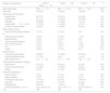
ResultsBaseline characteristicsThe study group consisted of 149 patients referred for coronary angiography, including 77 patients who previously underwent a SPECT-MPI, 39 who underwent DSE and 33 who underwent CTCA. The median time from a test result to coronary angiography was 3.5 (1.2–8.6) months. Characteristics of the study cohort stratified by the diagnostic test used are shown in Table 1. Patients undergoing SPECT-MP, DSE and CTCA were generally similar in terms of demographics and comorbidities, except for lower prevalence of CKD in CTCA and higher prevalence of previous revascularization in DSE. The CTCA group had a higher prevalence of patients with non-anginal chest pain, and fewer asymptomatic patients or those with dyspnea. Patients who underwent CTCA were less medicated with beta-blockers compared to patients who underwent the other non-invasive tests. All patients in the CTCA group received nitrate as part of the study protocol. Analysis of the baseline ECGs showed that LBBB was the most prevalent in all groups, compared to right ventricular pacing, and there were no significant differences in QRS duration between the groups.
Baseline characteristics of patients.
| Patients’ characteristics | SPECT-MPI(n=77) | DSE(n=39) | CTCA(n=33) | P |
|---|---|---|---|---|
| Age, years, mean | 66.6±11.0 | 68.4±10.5 | 65.0±10.2 | .360 |
| Sex, male | 41 (53.2) | 27 (69.2) | 15 (45.5) | .106 |
| Cardiovascular risk factors | ||||
| Hypertension | 56 (72.7) | 32 (82.1) | 21 (63.6) | .212 |
| Dyslipidemia | 55 (71.4) | 30 (76.9) | 25 (75.8) | .784 |
| Diabetes | 26 (33.8) | 17 (43.6) | 7 (21.2) | .134 |
| Obesity (BMI>30kg/m2) | 14 (18.2) | 11 (28.9) | 4 (12.1) | .185 |
| Current or former smoker | 19 (24.7) | 12 (30.8) | 8 (24.2) | .748 |
| Other medical history | ||||
| Chronic kidney disease (stage 3–5) | 10 (13) | 6 (15.4) | 0 (0) | .072 |
| Obstructive sleep apnea | 5 (6.5) | 4 (10.3) | 1 (3.0) | .472 |
| Atrial fibrillation | 4 (5.2) | 4 (10.3) | 0 (0) | .156 |
| Cerebrovascular disease | 5 (6.5) | 2 (5.1) | 0 (0) | .333 |
| Prior revascularization | 7 (9.1) | 9 (23.1) | 1 (3.0) | <.001 |
| PCI | 3 (2.1) | 2 (1.4) | 1 (3.0) | .951 |
| CABG | 4 (5.2) | 7 (17.9) | 0 (0) | .015 |
| Baseline ECG | ||||
| Left bundle branch block | 66 (85.7) | 37 (94.9) | 26 (78.8) | .130 |
| Ventricular paced rhythm | 11 (14.3) | 2 (5.1) | 7 (21.2) | .130 |
| QRS duration (ms) | 159±19.9 | 151±18.6 | 158±15.4 | .110 |
| Anti-ischemic medication at testing | ||||
| Beta-blocker | 47 (61.0) | 32 (82.1) | 9 (52.9) | .036 |
| Calcium channel blocker | 14 (18.2) | 12 (30.8) | 6 (35.3) | .166 |
| Nitrate | 2 (2.6) | 7 (17.9) | 33 (100)* | <.001 |
| Symptom classification | ||||
| Typical angina | 4 (5.2) | 4 (10.5) | 1 (3.0) | .376 |
| Atypical angina | 6 (7.8) | 4 (10.5) | 4 (2.1) | .751 |
| Non-anginal chest pain | 11 (14.3) | 6 (15.8) | 19 (57.6) | <.001 |
| Dyspnea | 33 (42.9) | 12 (31.6) | 6 (18.9) | .040 |
| Asymptomatic | 29 (41.4) | 17 (44.7) | 5 (15.2) | .015 |
| Pre-test probability (<5, 5–15, >15) | 9 (12), 36 (47), 27 (35) | 2 (5), 16 (41), 15 (38) | 5 (15), 15 (46), 13 (39) | .130 |
Data are expressed as no. (%) or mean±standard deviation. BMI, body mass index; CABG, coronary artery bypass grafting; CTCA, computed tomographic coronary angiography; DSE, dobutamine stress echocardiography; ECG, electrocardiogram; PCI, percutaneous coronary intervention; SPECT-MPI, single photon emission computed tomography myocardial perfusion imaging.
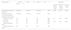
The overall results of SPECT-MPI, DSE, CTCA and coronary angiography are shown in Table 2. The prevalence of obstructive CAD in the study group was 30%, with no significant differences in the three cohorts (30%, 33% and 27% for SPECT-MPI, DSE and CTCA, respectively, P=.852). Despite the similar prevalence of obstructive CAD, there was a higher proportion of abnormal results in the SPECT-MPI cohort compared to the other cohorts (84% vs 64% vs 61% for SPECT-MPI, DSE and CTCA respectively, P=.009). There were no statistically significant differences between the prevalence of abnormal results in DSE and CTCA. Regarding the results of the diagnostic accuracy tests, specificity was found to be significantly lower in the SPECT-MPI group compared to the DSE and CTCA cohorts (18% vs 50% vs 54%, respectively, P=.012), driven by the low rate of truly negative results. Comparing the DSE and the CTCA cohorts, there were no significant differences in specificity between the two groups. There were no significant differences in sensitivity, negative predictive value and positive predictive value between the three groups.
Results of coronary angiography and diagnostic performance of SPECT-MPI, DSE and CTCA.
| Characteristic | SPECT-MPI(n=77) | DSE(n=39) | CTCA(n=33) | P | Adjusted P | ||
|---|---|---|---|---|---|---|---|
| SPECT-MPI vs DSE | SPECT-MPI vs CTCA | DSE vs CTCA | |||||
| Abnormal non-invasive test result | 65 (84.4) | 25 (64.1) | 20 (60.6) | .009* | .039* | .018* | >.999 |
| Obstructive CAD on coronary angiography | 23 (29.9) | 13 (33.3) | 9 (27.3) | .852 | |||
| Diagnostic performance | |||||||
| Predictive accuracy | 39 | 64 | 67 | .006* | .03* | .024* | >.999 |
| Sensitivity | 91 | 92 | 100 | .665 | |||
| Specificity | 18 | 50 | 54 | .002* | .012* | .003* | >.999 |
| Positive predictive value | 31 | 48 | 45 | .233 | |||
| Negative predictive value | 83 | 93 | 100 | .294 | |||
Data for exam results are shown as n (%). Diagnostic accuracy results are shown as %. CAD, coronary artery disease; CTCA, computed tomographic coronary angiography; DSE, dobutamine stress echocardiography; SPECT-MPI, single photon emission computed tomography myocardial perfusion imaging. P adjusted with Bonferroni correction.
Predicted accuracy was significantly lower in the SPECT-MPI group (39% vs 64% vs 67%, P=.006). When directly comparing the accuracy of the non-invasive tests, SPECT-MPI performance was lower than DSE (39% vs 64%, P=.03) and lower than CTCA (39% vs 67%, P=.024). The DSE performance was non-inferior to CTCA regarding predictive accuracy (64% vs 67%, P>.999).
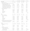
Diagnostic test characteristics and false positive analysisThe individual test results in the study group and the characterization of true and false positive results are shown in Table 3. In the SPECT-MPI cohort, the mean LVEF estimated by the study was 43±14%. Most patients (83%) had a partial or completely reversible perfusion defect. The most frequent location of the perfusion defect was in the apical segments (83%), followed by the septal segments (79%). The prevalence of septal defects was similar in patients with true positive and false positive results (85% vs 89%, P=.420). The same was true for apical defects (95% vs 87%, P=.655) suggesting that defect location is not a reliable marker to differentiate false positive results in these patients. Additionally, there were no differences between true and false positive results regarding the extent of the stress defect (P=.357) and the severity of the stress defect, assessed by the summed stress score (P=.789), the summed rest score (P=.793) or the summed difference score (P=.865).
Results of SPECT-MPI, DSE, and CTCA and characterization of true and false positive results.
| Total | True Positives | False positives | p value | |
|---|---|---|---|---|
| SPECT-MPI | N=77 | N=20 | N=45 | |
| LVEF (%, mean (SD)) | 42.7 (± 13.8) | 37.4 (± 13.0) | 44.1 (± 14.4) | 0.086 |
| Perfusion defect reversibility and segmental scoring | ||||
| Irreversible perfusion defect | 7 (9%) | 3 (15%) | 1 (2%) | 0.083 |
| Partially reversible perfusion defect | 34 (44%) | 11 (55%) | 22 (49%) | 0.789 |
| Completely reversible perfusion defect | 30 (39%) | 6 (30%) | 21 (47%) | 0.169 |
| Stress defect extent (%, median (Q1-Q3)) | 8 (5 – 15) | 12 (7 – 22) | 10 (5 – 16) | 0.357 |
| Summed stress score (median (Q1-Q3)) | 7 (4 – 11) | 7 (5 – 16) | 8 (5 – 11) | 0.789 |
| Summed rest score (median (Q1-Q3)) | 2 (0 – 4) | 3 (1 – 9) | 2 (1 – 4) | 0.793 |
| Summed difference score [mean (SD)] | 4.7 (± 3.2) | 5.1 (± 3.3) | 5.4 (± 2.9) | 0.865 |
| Perfusion defect location | ||||
| Septal | 61 (79%) | 17 (85%) | 40 (89%) | 0.420 |
| Anterior | 22 (29%) | 9 (45%) | 11 (24%) | 0.145 |
| Apical | 64 (83%) | 19 (95%) | 39 (87%) | 0.655 |
| Anterolateral | 9 (12%) | 4 (20%) | 3 (7%) | 0.200 |
| Inferolateral | 1 (1%) | 0 (0%) | 1 (2%) | - |
| Inferior | 10 (13%) | 3 (15%) | 7 (16%) | >0.999 |
| DSE | N=39 | N=12 | N=13 | |
| LVEF (%, mean (SD)) | 42.3 (± 13.0) | 39.8 (± 11.5) | 41.1 (± 13.6) | 0.795 |
| Location of ischemia/scar | ||||
| Septal | 19 (49%) | 10 (83%) | 9 (69%) | 0.645 |
| Anterior | 15 (39%) | 8 (67%) | 7 (54%) | 0.688 |
| Apical | 15 (39%) | 8 (67%) | 7 (54%) | 0.688 |
| Anterolateral | 14 (36%) | 8 (67%) | 6 (46%) | 0.302 |
| Inferolateral | 15 (39%) | 9 (75%) | 6 (46%) | 0.226 |
| Inferior | 18 (46%) | 9 (69%) | 9 (69%) | >0.999 |
| Ventricular arrhythmias reported during exam | ||||
| Frequent premature ventricular complexes | 17 (44%) | 7 (58%) | 7 (54%) | 0.821 |
| Non-sustained ventricular tachycardia | 5 (13%) | 0 (0%) | 2 (23%) | 0.076 |
| CTCA | N=33 | N=9 | N=11 | |
| Agatston score (median (IQ)) | 171 (45 – 425) | 373 (152-812) | 217 (125 – 465) | 0.408 |
| CAD-RADS | ||||
| 0-2 | 13 (39%) | 0 (0%) | 0 (0%) | - |
| 3 | 15 (46%) | 5 (55%) | 10 (91%) | 0.127 |
| 4 A-B | 5 (15%) | 4 (45%) | 1 (9%) | 0.127 |
| Obstructive coronary arteries | ||||
| Left main coronary artery | 0 (0%) | 0 (0%) | 0 (0%) | - |
| Left anterior descending artery | 20 (61%) | 9 (100%) | 9 (82%) | 0.479 |
| Left circumflex artery | 5 (15%) | 2 (22%) | 3 (27%) | >0.999 |
| Right coronary artery | 3 (9%) | 2 (22%) | 1 (9%) | 0.566 |
Data are expressed as no. (%), mean±standard deviation or median [interquartile range]. CAD-RADS, coronary artery disease-reporting and data system; CTCA, computed tomographic coronary angiography; DSE, dobutamine stress echocardiography; LVEF, left ventricular ejection fraction; SPECT-MPI, single photon emission computed tomography myocardial perfusion imaging.
In the DSE cohort, the mean LVEF was 42±13%. The most frequent location of ischemia or scar was in the septal wall (49%), followed by the inferior wall (46%). There were no differences in the location of ischemia or scar between true positives and false positives. Non-sustained ventricular tachycardia was an unreliable marker to differentiate true positive results (P=.076).
In the CTCA cohort, the median Agatston calcium score was 171 (45–425), with no differences between true and false positive results (P=.408). The lesion severity was also an unreliable predictor of false positive results since there were no differences between true and false positive results in patients with a CAD-RADS classification of 3 (P=.127) or 4 (P=.127). Similarly, the coronary artery involved was also an unreliable predictor of false positive results, with a similar distribution between the two groups for the anterior descending artery (P=.479), circumflex artery (P>.999) and right coronary artery (P=.566).
DiscussionThe authors present a retrospective study that directly compares the diagnostic performance of non-invasive anatomical and functional tests in the assessment of obstructive coronary artery disease in patients with LBBB and RVP. To this purpose, patients who underwent SPECT-MPI, DSE and CTCA were included. All patients underwent subsequent coronary angiography as a gold-standard test. Both DSE and CTCA were more accurate than SPECT in this cohort of patients, which was driven by a greater false positive rate (58%) in the SPECT cohort. Despite some baseline heterogeneity, the similar prevalence of significant CAD between the groups reinforces this finding, since a similar rate of abnormal results would be expected. Since the diagnostic performance of the DES was non-inferior to the CTCA, our findings suggest that the limitations of SPECT-MPI in patients with LBBB and RVP are not necessarily extended to other functional tests. Thus, an anatomical-based approach does not appear to be superior to a functional-based approach for the diagnosis of significant CAD in this group of patients. To our knowledge, this is the first comparative study of the performance of SPECT-MPI, DSE and CTCA in patients with LBBB and RVP.
Our findings are in agreement with previous studies citing false-positive septal perfusion defects in LBBB patients undergoing SPECT-MPI. A meta-analysis has reported a sensitivity and specificity in these patients of 89% and 41%, respectively,8 which is far inferior to that reported for the general population by previous meta-analyses (sensitivity ranging from 61 to 85% and specificity 79 to 85%).9,10 However, in our study, the specificity of SPECT-MPI was lower (18%). Considering the high false-positive rate of SPECT-MPI, the low referral rate for coronary angiography of patients with negative tests reduces the specificity of the test, as not all true negatives are included. Specifically in our study, only 10 patients with negative SPECT-MPI underwent coronary angiography, since patients who did not undergo coronary angiography were excluded.
The reduced specificity of SPECT-MPI is mostly explained by the high incidence of LBBB/RVP-related septal and apical perfusion defects.5 Several non-CAD mechanisms have been proposed for these findings, including decreased septal perfusion due to lower oxygen demand caused by less myocardial work in this region, diastolic compression of perforator branches due to asynchronous contraction, microvascular dysfunction and septal fibrosis in patients with cardiomyopathic changes.2,3 The perfusion defect may also be purely artifactual. In the semi-quantitative perfusion analysis by pixel-measured counts, there is an assumption of similar systolic thickness across LV walls. LBBB patients, however, usually have asymmetric thickening of the lateral systolic wall relative to the septum, which may mimic perfusion heterogeneity by a so-called partial volume effect.2 Although some authors describe mechanisms to distinguish true and false positive results,3 this is often difficult in real-world practice. Our study found no significant differences between true or false positive results regarding LVEF, perfusion defect location, reversibility, extent or intensity of the defect.
The results of studies evaluating the diagnostic accuracy of DSE in LBBB have been so far controversial. Some authors report poor specificity (64%),11 while other researchers argue that in experienced hands it may reach a sensitivity and specificity exceeding 90% and a better performance than SPECT-MPI in the diagnosis of significant CAD in the left coronary artery territory.12 Our results support the role of DSE in diagnosing significant CAD in these patients, with a high sensitivity but a specificity of 50%, lower than reported. Although accurate detection of regional wall motion abnormalities may be challenging in these patients, myocardial thickening remains nearly normal in the absence of significant CAD. Therefore, experienced operators may identify an ischemic response by stress-induced wall thickening impairment, including failure to improve wall thickening relative to the hyperkinetic response to maximal stress.13
The frequency of referral of LBBB patients for coronary angiography due to abnormal functional tests suggests a potential role for anatomical tests in the assessment of significant CAD in these patients. The fact that CTCA is not influenced by perfusion or wall motion artifacts makes it a promising non-invasive technique in these patients. This exam has previously been compared to coronary angiography in a study of 66 patients with LBBB, having shown excellent accuracy in detecting obstructive CAD, correctly assessing 95% of patients with significant CAD and identifying all the patients who needed revascularization.14 In our study, CTCA showed the highest sensitivity, specificity and predictive accuracy values, although with no statistically significant differences compared to DSE. It also identified all patients with an indication for revascularization. Despite the potential role of the CTCA, the known limitations of this test, such as coronary calcification, arrhythmias and chronic kidney disease also apply to these patients. This is a favorable point for DSE given that it is less affected by these conditions and is also non-inferior to CTCA in the study of these patients.
Study limitationsDue to the observational nature of the study, there is a risk of referral bias. Since patients with negative test results are not commonly referred to coronary angiography, this introduces referral bias, which has been shown to result in an overestimation of sensitivity and underestimation of specificity.15 In our study, this specificity bias may be more relevant concerning SPECT-MPI since few patients with a normal test underwent coronary angiography and also because there is a high rate of false negatives, reducing specificity.
The small sample size, especially in the DSE and CTCA groups, is also a limitation of the study. Additionally, since patients with previous myocardial infarction or incomplete revascularization were excluded, it may influence the external validity of the results.
The different segmentation of the heart adopted by non-invasive functional exams could be a source of bias and constitute a limitation of the study.
Given that the time between the non-invasive test and coronary angiography was not the same for all patients, this is also a limitation of the study. Furthermore, the gold standard in this analysis was coronary angiography without the use of invasive functional tests, which would have been the true gold standard of non-invasive functional tests.
Finally, the inclusion of patients who underwent myocardial perfusion assessment by magnetic resonance imaging was not possible because of the low referral of patients meeting inclusion criteria in the study centers.
ConclusionIn patients with LBBB and RVP, SPECT-MPI performed worse than DSE and CTCA for the diagnosis of significant CAD. CTCA and DSE show similar performance in the assessment of obstructive CAD in patients with LBBB and RVP, showing that both anatomical and functional tests have a role in the study of significant coronary disease in these patients.
- -
A significant number of patients with LBBB and RVP are referred for evaluation of significant coronary disease. In these patients, the use of functional tests can be challenging considering the artifacts associated with SPECT-MPI and abnormal septal motion secondary to LBBB that can affect DSE results.
- -
This is the first comparison of the performance of SPECT-MPI, DSE and CTCA in patients with LBBB and RVP.
- -
SPECT-MPI has an inferior performance compared to DSE and CTCA, with lower diagnostic accuracy.
- -
Despite abnormal septal movement secondary to LBBB, DSE has a diagnostic performance that is non-inferior to CTCA and should be considered in the study of significant CAD in patients with LBBB and RVP, if performed by experienced hands.
None declared.
Ethical considerationsThis study was approved by the local ethics committee. Informed consent was not required due to the observational nature of the study. The authors followed SAGER guidelines for addressing sex and gender variables.
Statement on the use of artificial intelligenceNo artificial intelligence was used.
Authors’ contributionsAll authors participated in the design of the protocol and analysis of data, the drafting the work and gave their final approval of the version to be published.
Conflicts of interestThere are no conflicts of interest.