
Chloroquine (CQ) and hydroxychloroquine (HCQ), initially developed for malaria prophylaxis, are now widely used in rheumatic diseases, such as systemic lupus erythematosus and rheumatoid arthritis. Despite their therapeutic benefits, these drugs can induce cardiotoxicity, particularly cardiomyopathy. This systematic review evaluates the prevalence, clinical features, and diagnostic challenges of CQ/HCQ-induced cardiomyopathy.
MethodsA comprehensive literature search was conducted in PubMed, EMBASE, and Cochrane databases, identifying 60 studies reporting 79 cases of CQ/HCQ-induced cardiomyopathy. Inclusion criteria required histopathological confirmation via endomyocardial biopsy. Data on clinical presentations, diagnostic findings, and outcomes were extracted and analyzed.
ResultsPatients were predominantly female (76%), with a median age of 56 years. The cardiomyopathies observed included hypertrophic, dilated, and restrictive forms, frequently presenting with heart failure and conduction disorders, such as atrioventricular block and arrhythmias. Cardiac magnetic resonance imaging and endomyocardial biopsy revealed characteristic toxicological changes, including myocyte vacuolization and fibrosis. Management often involved discontinuation of therapy, but severe cases progressed to cardiac transplantation or resulted in mortality (19%).
ConclusionsCQ/HCQ-induced cardiomyopathy is an underrecognized complication with significant morbidity and mortality. Early detection through continuous cardiac monitoring in patients on long-term CQ/HCQ therapy is crucial. Effective management, including timely drug discontinuation, may improve outcomes, although prognosis remains guarded in advanced stages.
La cloroquina (CQ) y la hidroxicloroquina (HCQ), desarrolladas inicialmente para la profilaxis de la malaria, son hoy en día ampliamente utilizadas para el tratamiento de algunas enfermedades reumáticas, como el lupus eritematoso sistémico y la artritis reumatoide. A pesar de sus beneficios terapéuticos, estos fármacos pueden ocasionar toxicidad cardiovascular, concretamente cardiomiopatía. Esta revisión sistemática evalúa la prevalencia, las características clínicas y los retos diagnósticos de la cardiomiopatía inducida por CQ/HCQ.
MétodosSe realizó una búsqueda exhaustiva en las bases de datos PubMed, EMBASE y Cochrane, identificando 60 estudios que comunican 79 casos de cardiomiopatía inducida por CQ/HCQ. Los criterios de inclusión requerían la confirmación histopatológica mediante biopsia endomiocárdica. Se extrajeron y analizaron datos sobre presentación clínica, hallazgos diagnósticos y resultados.
ResultadosLos pacientes fueron predominantemente mujeres (76%), con una edad media de 56 años. Las cardiomiopatías observadas incluyeron formas hipertróficas, dilatadas y restrictivas, que frecuentemente se presentaron con insuficiencia cardiaca y trastornos de la conducción, como bloqueo auriculoventricular y arritmias. La resonancia magnética cardiaca y la biopsia endomiocárdica revelaron cambios tóxicos característicos, como vacuolización de miocitos y fibrosis. El manejo generalmente requirió la suspensión del tratamiento, y algunos casos graves progresaron a trasplante cardiaco o resultaron en mortalidad (19%).
ConclusionesLa cardiomiopatía inducida por CQ/HCQ es una complicación infradiagnosticada con una morbimortalidad significativa. La detección precoz mediante seguimiento cardiológico cercano en pacientes que reciben tratamiento prolongado con CQ/HCQ es crucial. Un tratamiento efectivo, incluyendo la suspensión oportuna del fármaco, puede mejorar los resultados, aunque el pronóstico sigue siendo complejo en etapas avanzadas.
Chloroquine (CQ) was first discovered in 1934 and has been widely used for malaria prevention since World War II.1 A decade later, CQ and hydroxychloroquine (HCQ) were introduced for the treatment of rheumatic diseases and are now extensively used in managing conditions such as systemic lupus erythematosus (SLE), rheumatoid arthritis (RA), and other connective tissue disorders.2 Despite their therapeutic efficacy, both drugs have been increasingly associated with significant toxicities, particularly affecting the cardiovascular system.
HCQ differs structurally from CQ due to a hydroxyl group attached to the side chain's ethyl group, which contributes to its lower toxicity profile.1 Both medications are rapidly absorbed, with tissue concentrations reaching a steady state within 1–3 months (corresponding to five half-lives). Upon discontinuation, tissue concentrations decrease by 90–95% within 15 days.3,4 Although HCQ is considered to have a lower risk of toxicity, it has been implicated in a range of dose-dependent adverse effects, particularly when used in long-term therapy.1
Among the various toxicities, cardiac toxicity represents a serious and often underdiagnosed complication of long-term CQ and HCQ therapy. The primary mechanisms of cardiotoxicity involve lysosomal enzyme dysfunction, which leads to impaired degradation of intracellular materials and the accumulation of metabolic byproducts such as glycogen and phospholipids within cardiomyocytes.4,5 This disruption causes cellular vacuolization, hypertrophy, and fibrosis, culminating in cardiomyopathy and conduction disturbances.6
Cardiotoxicity from CQ/HCQ manifests as conduction disorders, including atrioventricular block (AVB), and progressive forms of cardiomyopathy that can lead to severe heart failure (HF). Despite the severe implications for morbidity and mortality, this form of cardiotoxicity remains underrecognized in clinical practice.6
Given the potential for severe cardiac complications, a comprehensive understanding of the toxicological mechanisms underlying CQ/HCQ-induced cardiotoxicity is essential for early detection and prevention. This review aims to explore the prevalence, clinical manifestations, diagnostic strategies, and management of CQ/HCQ-induced cardiomyopathy, with a focus on elucidating the toxicological pathways involved. By enhancing awareness and understanding of these complications, we can improve outcomes for patients undergoing long-term therapy with these drugs.
MethodsProtocol and literature searchWe conducted a comprehensive literature search to investigate the association between CQ or HCQ and cardiomyopathy, particularly focusing on their toxicological effects on the cardiovascular system. The search, conducted via PubMed, EMBASE and Cochrane up to June 2024, employed a combination of keywords and Medical Subject Headings (MeSH) terms: “chloroquine” OR “hydroxychloroquine” AND “cardiomyopathy” OR “cardiomyopathies”. Our review follows PRISMA guidelines, ensuring transparency and reproducibility.
Eligibility criteriaStudies were included if they met the following criteria: (a) discussed the association between CQ or HCQ and cardiomyopathy and (b) confirmed the diagnosis of CQ/HCQ-induced cardiomyopathy through endomyocardial biopsy. Exclusion criteria included duplicate publications, unavailable full texts, studies unrelated to CQ/HCQ cardiotoxicity, or those not involving histopathological confirmation of cardiomyopathy.
Data collection and managementTwo authors (V.D. Neto, J.G. Fiúza) independently screened all titles and abstracts for relevance, followed by a full-text review of eligible articles. Data on study populations, clinical characteristics, drug dosage, toxicological findings, and patient outcomes were extracted to ensure the inclusion of the most relevant and up-to-date information. Discrepancies in data extraction or study eligibility were resolved by discussion or consultation with a third reviewer. This process ensured consistency and accuracy across all reviewed studies.
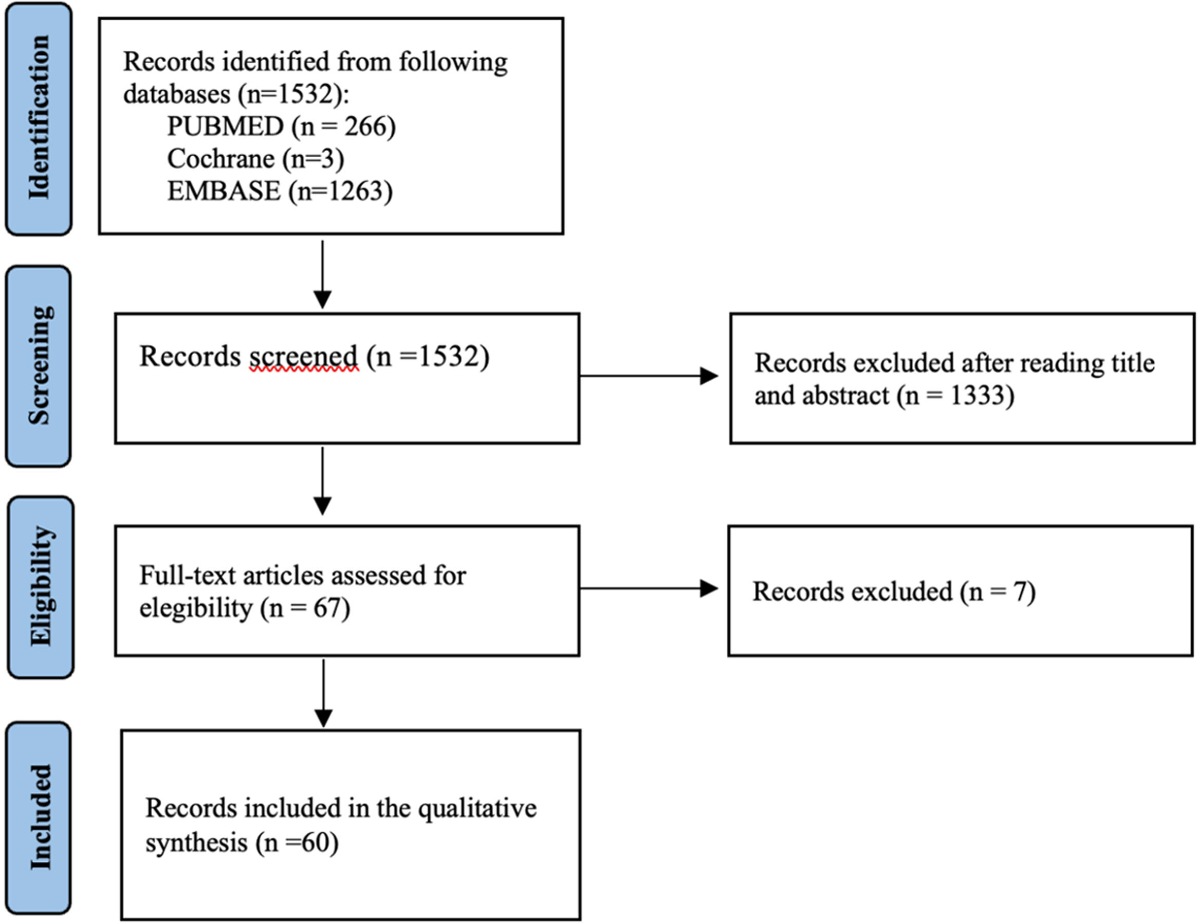
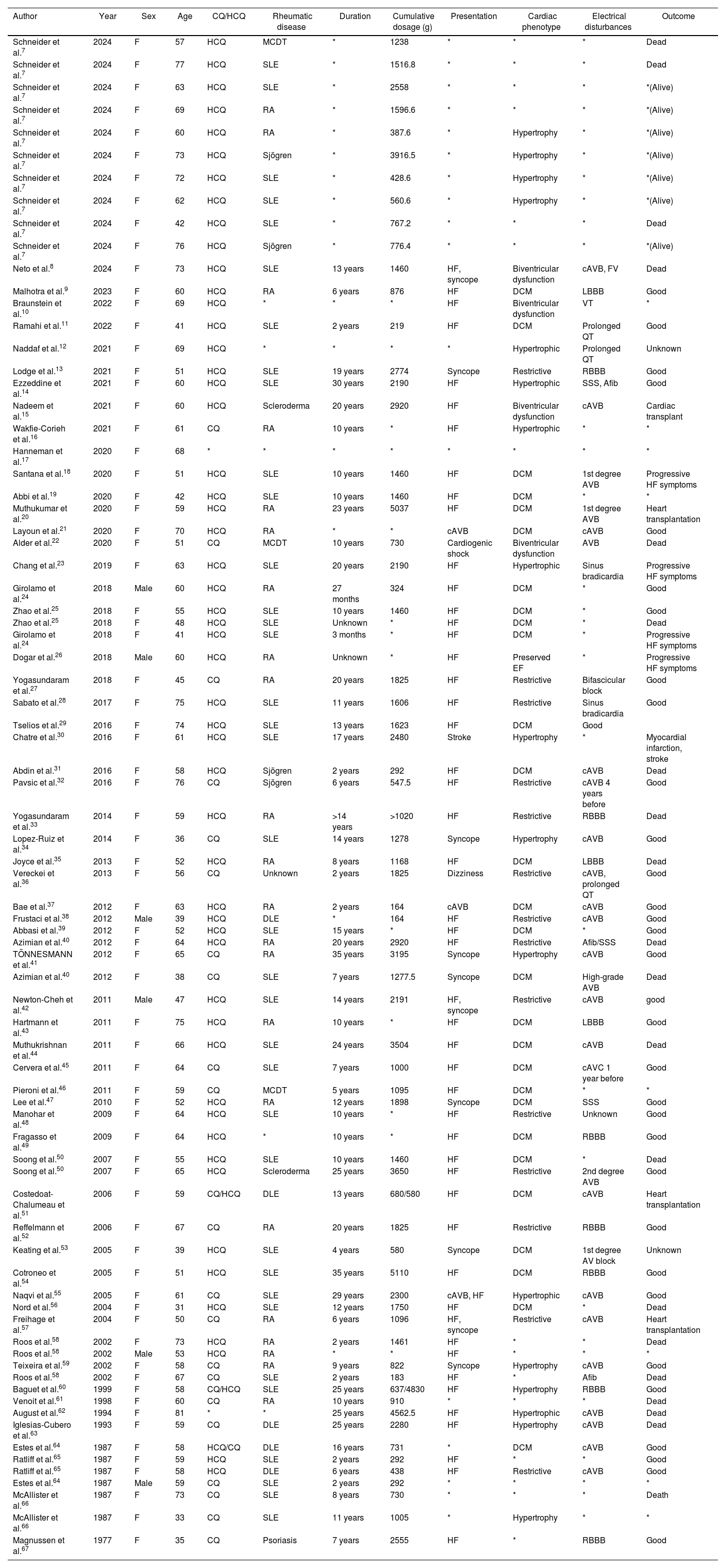
Search resultsOur search yielded 266 records. After screening the titles and abstracts, 199 articles were excluded for not meeting the inclusion criteria, leaving 67 for full-text review. A total of 60 articles were included in the final qualitative synthesis, documenting 79 cases of CQ/HCQ-induced cardiomyopathy. Cases are described in more detail in Table 1. Most studies were individual case reports or small case series, with no randomized controlled trials identified. A PRISMA flowchart (Fig. 1) details the selection process.
Information about clinical cases included.
| Author | Year | Sex | Age | CQ/HCQ | Rheumatic disease | Duration | Cumulative dosage (g) | Presentation | Cardiac phenotype | Electrical disturbances | Outcome |
|---|---|---|---|---|---|---|---|---|---|---|---|
| Schneider et al.7 | 2024 | F | 57 | HCQ | MCDT | * | 1238 | * | * | * | Dead |
| Schneider et al.7 | 2024 | F | 77 | HCQ | SLE | * | 1516.8 | * | * | * | Dead |
| Schneider et al.7 | 2024 | F | 63 | HCQ | SLE | * | 2558 | * | * | * | *(Alive) |
| Schneider et al.7 | 2024 | F | 69 | HCQ | RA | * | 1596.6 | * | * | * | *(Alive) |
| Schneider et al.7 | 2024 | F | 60 | HCQ | RA | * | 387.6 | * | Hypertrophy | * | *(Alive) |
| Schneider et al.7 | 2024 | F | 73 | HCQ | Sjögren | * | 3916.5 | * | Hypertrophy | * | *(Alive) |
| Schneider et al.7 | 2024 | F | 72 | HCQ | SLE | * | 428.6 | * | Hypertrophy | * | *(Alive) |
| Schneider et al.7 | 2024 | F | 62 | HCQ | SLE | * | 560.6 | * | Hypertrophy | * | *(Alive) |
| Schneider et al.7 | 2024 | F | 42 | HCQ | SLE | * | 767.2 | * | * | * | Dead |
| Schneider et al.7 | 2024 | F | 76 | HCQ | Sjögren | * | 776.4 | * | * | * | *(Alive) |
| Neto et al.8 | 2024 | F | 73 | HCQ | SLE | 13 years | 1460 | HF, syncope | Biventricular dysfunction | cAVB, FV | Dead |
| Malhotra et al.9 | 2023 | F | 60 | HCQ | RA | 6 years | 876 | HF | DCM | LBBB | Good |
| Braunstein et al.10 | 2022 | F | 69 | HCQ | * | * | * | HF | Biventricular dysfunction | VT | * |
| Ramahi et al.11 | 2022 | F | 41 | HCQ | SLE | 2 years | 219 | HF | DCM | Prolonged QT | Good |
| Naddaf et al.12 | 2021 | F | 69 | HCQ | * | * | * | * | Hypertrophic | Prolonged QT | Unknown |
| Lodge et al.13 | 2021 | F | 51 | HCQ | SLE | 19 years | 2774 | Syncope | Restrictive | RBBB | Good |
| Ezzeddine et al.14 | 2021 | F | 60 | HCQ | SLE | 30 years | 2190 | HF | Hypertrophic | SSS, Afib | Good |
| Nadeem et al.15 | 2021 | F | 60 | HCQ | Scleroderma | 20 years | 2920 | HF | Biventricular dysfunction | cAVB | Cardiac transplant |
| Wakfie-Corieh et al.16 | 2021 | F | 61 | CQ | RA | 10 years | * | HF | Hypertrophic | * | * |
| Hanneman et al.17 | 2020 | F | 68 | * | * | * | * | * | * | * | * |
| Santana et al.18 | 2020 | F | 51 | HCQ | SLE | 10 years | 1460 | HF | DCM | 1st degree AVB | Progressive HF symptoms |
| Abbi et al.19 | 2020 | F | 42 | HCQ | SLE | 10 years | 1460 | HF | DCM | * | * |
| Muthukumar et al.20 | 2020 | F | 59 | HCQ | RA | 23 years | 5037 | HF | DCM | 1st degree AVB | Heart transplantation |
| Layoun et al.21 | 2020 | F | 70 | HCQ | RA | * | * | cAVB | DCM | cAVB | Good |
| Alder et al.22 | 2020 | F | 51 | CQ | MCDT | 10 years | 730 | Cardiogenic shock | Biventricular dysfunction | AVB | Dead |
| Chang et al.23 | 2019 | F | 63 | HCQ | SLE | 20 years | 2190 | HF | Hypertrophic | Sinus bradicardia | Progressive HF symptoms |
| Girolamo et al.24 | 2018 | Male | 60 | HCQ | RA | 27 months | 324 | HF | DCM | * | Good |
| Zhao et al.25 | 2018 | F | 55 | HCQ | SLE | 10 years | 1460 | HF | DCM | * | Good |
| Zhao et al.25 | 2018 | F | 48 | HCQ | SLE | Unknown | * | HF | DCM | * | Dead |
| Girolamo et al.24 | 2018 | F | 41 | HCQ | SLE | 3 months | * | HF | DCM | * | Progressive HF symptoms |
| Dogar et al.26 | 2018 | Male | 60 | HCQ | RA | Unknown | * | HF | Preserved EF | * | Progressive HF symptoms |
| Yogasundaram et al.27 | 2018 | F | 45 | CQ | RA | 20 years | 1825 | HF | Restrictive | Bifascicular block | Good |
| Sabato et al.28 | 2017 | F | 75 | HCQ | SLE | 11 years | 1606 | HF | Restrictive | Sinus bradicardia | Good |
| Tselios et al.29 | 2016 | F | 74 | HCQ | SLE | 13 years | 1623 | HF | DCM | Good | |
| Chatre et al.30 | 2016 | F | 61 | HCQ | SLE | 17 years | 2480 | Stroke | Hypertrophy | * | Myocardial infarction, stroke |
| Abdin et al.31 | 2016 | F | 58 | HCQ | Sjögren | 2 years | 292 | HF | DCM | cAVB | Dead |
| Pavsic et al.32 | 2016 | F | 76 | CQ | Sjögren | 6 years | 547.5 | HF | Restrictive | cAVB 4 years before | Good |
| Yogasundaram et al.33 | 2014 | F | 59 | HCQ | RA | >14 years | >1020 | HF | Restrictive | RBBB | Dead |
| Lopez-Ruiz et al.34 | 2014 | F | 36 | CQ | SLE | 14 years | 1278 | Syncope | Hypertrophy | cAVB | Good |
| Joyce et al.35 | 2013 | F | 52 | HCQ | RA | 8 years | 1168 | HF | DCM | LBBB | Dead |
| Vereckei et al.36 | 2013 | F | 56 | CQ | Unknown | 2 years | 1825 | Dizziness | Restrictive | cAVB, prolonged QT | Good |
| Bae et al.37 | 2012 | F | 63 | HCQ | RA | 2 years | 164 | cAVB | DCM | cAVB | Good |
| Frustaci et al.38 | 2012 | Male | 39 | HCQ | DLE | * | 164 | HF | Restrictive | cAVB | Good |
| Abbasi et al.39 | 2012 | F | 52 | HCQ | SLE | 15 years | * | HF | DCM | * | Good |
| Azimian et al.40 | 2012 | F | 64 | HCQ | RA | 20 years | 2920 | HF | Restrictive | Afib/SSS | Dead |
| TÖNNESMANN et al.41 | 2012 | F | 65 | CQ | RA | 35 years | 3195 | Syncope | Hypertrophy | cAVB | Good |
| Azimian et al.40 | 2012 | F | 38 | CQ | SLE | 7 years | 1277.5 | Syncope | DCM | High-grade AVB | Dead |
| Newton-Cheh et al.42 | 2011 | Male | 47 | HCQ | SLE | 14 years | 2191 | HF, syncope | Restrictive | cAVB | good |
| Hartmann et al.43 | 2011 | F | 75 | HCQ | RA | 10 years | * | HF | DCM | LBBB | Good |
| Muthukrishnan et al.44 | 2011 | F | 66 | HCQ | SLE | 24 years | 3504 | HF | DCM | cAVB | Dead |
| Cervera et al.45 | 2011 | F | 64 | CQ | SLE | 7 years | 1000 | HF | DCM | cAVC 1 year before | Good |
| Pieroni et al.46 | 2011 | F | 59 | CQ | MCDT | 5 years | 1095 | HF | DCM | * | * |
| Lee et al.47 | 2010 | F | 52 | HCQ | RA | 12 years | 1898 | Syncope | DCM | SSS | Good |
| Manohar et al.48 | 2009 | F | 64 | HCQ | SLE | 10 years | * | HF | Restrictive | Unknown | Good |
| Fragasso et al.49 | 2009 | F | 64 | HCQ | * | 10 years | * | HF | DCM | RBBB | Good |
| Soong et al.50 | 2007 | F | 55 | HCQ | SLE | 10 years | 1460 | HF | DCM | * | Dead |
| Soong et al.50 | 2007 | F | 65 | HCQ | Scleroderma | 25 years | 3650 | HF | Restrictive | 2nd degree AVB | Good |
| Costedoat-Chalumeau et al.51 | 2006 | F | 59 | CQ/HCQ | DLE | 13 years | 680/580 | HF | DCM | cAVB | Heart transplantation |
| Reffelmann et al.52 | 2006 | F | 67 | CQ | RA | 20 years | 1825 | HF | Restrictive | RBBB | Good |
| Keating et al.53 | 2005 | F | 39 | HCQ | SLE | 4 years | 580 | Syncope | DCM | 1st degree AV block | Unknown |
| Cotroneo et al.54 | 2005 | F | 51 | HCQ | SLE | 35 years | 5110 | HF | DCM | RBBB | Good |
| Naqvi et al.55 | 2005 | F | 61 | CQ | SLE | 29 years | 2300 | cAVB, HF | Hypertrophic | cAVB | Good |
| Nord et al.56 | 2004 | F | 31 | HCQ | SLE | 12 years | 1750 | HF | DCM | * | Dead |
| Freihage et al.57 | 2004 | F | 50 | CQ | RA | 6 years | 1096 | HF, syncope | Restrictive | cAVB | Heart transplantation |
| Roos et al.58 | 2002 | F | 73 | HCQ | RA | 2 years | 1461 | HF | * | * | Dead |
| Roos et al.58 | 2002 | Male | 53 | HCQ | RA | * | * | HF | * | * | * |
| Teixeira et al.59 | 2002 | F | 58 | CQ | RA | 9 years | 822 | Syncope | Hypertrophy | cAVB | Good |
| Roos et al.58 | 2002 | F | 67 | CQ | SLE | 2 years | 183 | HF | * | Afib | Dead |
| Baguet et al.60 | 1999 | F | 58 | CQ/HCQ | SLE | 25 years | 637/4830 | HF | Hypertrophy | RBBB | Good |
| Venoit et al.61 | 1998 | F | 60 | CQ | RA | 10 years | 910 | * | * | * | Dead |
| August et al.62 | 1994 | F | 81 | * | * | 25 years | 4562.5 | HF | Hypertrophic | cAVB | Dead |
| Iglesias-Cubero et al.63 | 1993 | F | 59 | CQ | DLE | 25 years | 2280 | HF | Hypertrophy | cAVB | Dead |
| Estes et al.64 | 1987 | F | 58 | HCQ/CQ | DLE | 16 years | 731 | * | DCM | cAVB | Good |
| Ratliff et al.65 | 1987 | F | 59 | HCQ | SLE | 2 years | 292 | HF | * | * | Good |
| Ratliff et al.65 | 1987 | F | 58 | HCQ | DLE | 6 years | 438 | HF | Restrictive | cAVB | Good |
| Estes et al.64 | 1987 | Male | 59 | CQ | SLE | 2 years | 292 | * | * | * | * |
| McAllister et al.66 | 1987 | F | 73 | CQ | SLE | 8 years | 730 | * | * | * | Death |
| McAllister et al.66 | 1987 | F | 33 | CQ | SLE | 11 years | 1005 | * | Hypertrophy | * | * |
| Magnussen et al.67 | 1977 | F | 35 | CQ | Psoriasis | 7 years | 2555 | HF | * | RBBB | Good |
Afib, atrial fibrillation; AVB, atrioventricular block; cAVB, complete atrioventricular block; CQ, chloroquine; DCM, dilated cardiomyopathy; DLE, discoid lupus erythematosus; F, female; HCQ, hydroxychloroquine; HF, heart failure; LBBB, left bundle branch block; MCDT, mixed connection tissue disorder; RA, rheumatoid arthritis; RBBB, right bundle branch block; SLE, systemic lupus erythematosus; SSS, sick sinus syndrome.
Most patients were female (n=76%), with a median age of 60 years (range: 35–81 years). This demographic is reflective of the higher prevalence of autoimmune diseases, such as SLE and RA, among women.
SLE was the most common underlying condition, present in 51% of cases (n=40), followed by RA in 25% of cases (n=20). Other conditions included Sjögren's syndrome (n=9, 11%) and mixed connective tissue disease (n=5, 6%).
Duration and cumulative dosage of therapyThe duration of CQ/HCQ therapy varied significantly across patients, ranging from 2 months to over 35 years (median duration: 10 years). The cumulative dosages ranged from 164g to 5110g, with a median cumulative dose of 1825g. Prolonged exposure and higher cumulative doses were associated with increased risk of cardiotoxicity, particularly in cases with durations exceeding 10 years of continuous therapy.
Cardiac manifestations: cardiomyopathyHF was the predominant clinical presentation, occurring in 62% of patients (n=49). HCQ-induced cardiomyopathy is predominantly described as hypertrophic cardiomyopathy (HCM); however, cases of dilated (DCM) and restrictive cardiomyopathy (RCM) have also been reported.
Concentric hypertrophy was the most frequently observed cardiac phenotype, present in 45% of cases (n=36). For cases with available data (n=13), the median cumulative dosage was 2190g (range: 387.6–3916.5g), with a median exposure duration of 20 years (range: 9–35 years).
The second most common phenotype was DCM identified in 35% of cases (n=28). For the cases with reported data (n=24), the median cumulative dosage was 1368.8g (range: 164–5110g), and the median exposure duration was 10 years (range: 0.25–35 years).
The least frequent phenotype was RCM, observed in 20% of cases (n=16). For the cases with dosage and duration data (n=12), the median cumulative dosage was 1825g (range 164–3650g), and the median exposure duration was 14 years (range 2–25 years).
HCM was the most common phenotype with apparently shorter therapy durations and a broader dosage range, indicating greater variability in the factors contributing to its development. HCM occurred after longer therapy durations (median 20 years) and moderately high cumulative dosages, suggesting that prolonged exposure may be a critical factor in its pathogenesis. RCM was intermediate between HCM and DCM in both therapy duration and cumulative dosage. This suggests that RCM might develop after prolonged therapy but potentially less so than HCM.
Patients with HCM had features comparable to those observed in other lysosomal storage disorders, such as Anderson-Fabry and Danon disease, which share a similar pathophysiology involving lysosomal dysfunction and intracellular storage of metabolic products.68,69
Hydroxychloroquine toxicity may lead to false-positive results in cardiac scintigraphy using bisphosphonates, potentially resulting in a misdiagnosis of cardiac amyloidosis. In such cases, endomyocardial biopsy plays a crucial role in confirming the diagnosis.16
Conduction disturbancesConduction abnormalities were present in 40% of cases (n=32), with the most common disturbances being AVB. Specifically, third-degree AVB occurred in 15% of cases (n=12), often requiring permanent pacemaker implantation. QT prolongation was identified in 10% of cases (n=8), which in some instances led to Torsades de Pointes and ventricular tachycardia, requiring implantable cardioverter-defibrillator placement.8,10
Cardiac magnetic resonance imaging (CMR)CMR has been instrumental in diagnosing and characterizing the extent of cardiomyopathy in patients with CQ HCQ-induced cardiomyopathy. Common findings include late gadolinium enhancement (LGE), indicative of myocardial fibrosis, and low native T1 mapping values, reflecting lipid overload associated with the lysosomal storage nature of the disease. However, in some cases, prolonged exposure to HCQ may lead to diffuse myocardial fibrosis and edema, resulting in strikingly increased native T1 values. This is likely due to the progression of the pathological process from glycosphingolipid accumulation to fibrosis and inflammation, as indicated by increased T1 and T2 values.7,8,15,33,70
The CMR findings in HCQ-induced cardiomyopathy closely resemble those observed in Anderson-Fabry disease (AFD), making differential diagnosis challenging. In such cases, additional diagnostic methods, such as genetic testing and endomyocardial biopsy, may be necessary to confirm the diagnosis.15,33,70
Both HCQ-induced cardiomyopathy and Anderson-Fabry disease often present with decreased T1 mapping values due to lipid or lysosomal product accumulation. However, in AFD, T1 values are typically markedly reduced (e.g., often below 900ms on 1.5T scanners), which can help differentiate the two conditions. In Danon disease, T1 values do not follow a consistent pattern but are often elevated due to extensive myocardial fibrosis.15,33,70
The patterns of LGE also vary between these conditions:
- -
HCQ-induced cardiomyopathy: Patchy, mid-myocardial, or subepicardial enhancement, consistent with non-ischemic fibrosis.
- -
Anderson-Fabry disease: LGE commonly involves the basal inferolateral wall of the left ventricle.
- -
Danon disease: LGE is often diffuse with extensive myocardial involvement, though typically non-specific. 15,33,70 T2 mapping may reveal subtle increases in HCQ-induced cardiomyopathy, reflecting inflammation or edema. Active Danon disease frequently exhibits higher T2 values due to pronounced inflammation. In contrast, early-stage Anderson-Fabry disease typically presents with normal T2 values, with elevations observed only in advanced stages of the disease.15,33,70
Endomyocardial biopsy (EMB) remains the definitive diagnostic tool for HCQ-induced cardiomyopathy, particularly in distinguishing it from other forms of cardiomyopathy and cardiac manifestations of connective tissue diseases.
Lysosomal storage disorders, including HCQ-induced cardiomyopathy, involve the accumulation of substances within lysosomes, leading to hypertrophy and vacuolization in cardiomyocytes. Myocyte vacuolization is a hallmark of drug-induced lysosomal storage disease. Focal interstitial fibrosis and myocyte hypertrophy further contribute to cardiac dysfunction.7–67
Although vacuolated cardiomyocytes visible under light microscopy are strongly suggestive of lysosomal storage disease, they are not specific to HCQ-induced cardiomyopathy and may also be seen in Anderson-Fabry and Danon diseases. Histological differences, such as the absence of periodic acid-Schiff (PAS) positivity in HCQ-induced cases, can help exclude glycogen storage disorders.7–67
Electron microscopy is considered necessary to confirm HCQ-induced cardiomyopathy when light microscopy findings are inconclusive. The presence of curvilinear bodies in biopsy specimens is pathognomonic for HCQ-induced cardiomyopathy, distinguishing it from Anderson-Fabry (characterized by globotriaosylceramide accumulation) and Danon disease.33 However, when electron microscopy is unavailable, clinical judgment and light microscopy findings may suffice for diagnosis in appropriate scenarios.8
Outcomes and prognosisHCQ-induced cardiomyopathy may show partial or complete reversibility after discontinuing the drug. This reversibility appears to depend on the severity of myocardial involvement and how early the condition is detected and treated. However, in cases of advanced cardiomyopathy with significant fibrosis, the damage may be irreversible, as fibrotic tissue does not recover.33
Despite discontinuation of CQ/HCQ therapy in most cases, the outcomes varied widely. Cardiac transplantation was required in 5% of cases (n=4), reflecting the severity of left ventricular dysfunction in certain patients.15,20,21 Mortality rate was 19% (n=15), with most deaths occurring within 1 year of diagnosis despite cessation of CQ/HCQ and initiation of aggressive HF treatment7,8,22,25,31 and 40% of patients (n=32) showed clinical improvement following cessation of therapy and appropriate heart failure management.
DiscussionThis systematic review highlights the significant risk of HCQ and CQ-induced cardiomyopathy in patients undergoing long-term treatment, particularly for SLE and RA. Our findings, drawn from 79 individual cases, reveal that both drugs can cause severe cardiac toxicity, manifesting as hypertrophic, dilated, and restrictive cardiomyopathies. While HCQ is generally regarded as having a more favorable safety profile compared to CQ, our review demonstrates that both drugs can lead to severe cardiac events, particularly after prolonged exposure or at high cumulative doses.1,3
HCQ and CQ belong to the category of cationic amphiphilic drugs, which preferentially bind to phospholipids and accumulate in lysosomes.3 This accumulation disrupts normal lysosomal function, leading to a rise in lysosomal pH and subsequent inhibition of lysosomal enzymes such as alpha- and beta-galactosidase. This enzyme inhibition impairs intracellular degradation and causes the accumulation of phospholipids and glycogen, mimicking the pathophysiology of lysosomal storage diseases.70,71 As these metabolic products accumulate within the cardiac myocytes, they result in vacuolated cytoplasm, myocyte hypertrophy, and eventually myocardial fibrosis.72
Patients has frequently associated diastolic dysfunction, and in many cases, the histopathological findings resembled those of genetic lysosomal storage disorders such as Anderson-Fabry and Danon diseases.68,69 This suggests that the lysosomal dysfunction caused by HCQ and CQ shares common pathways with these genetic conditions.
The accumulation of metabolic products also affects the cardiac conduction system, leading to a range of conduction disturbances. These conduction abnormalities often manifested as bradyarrhythmia requiring permanent pacemaker implantation or as life-threatening tachyarrhythmias, such as ventricular tachycardia, requiring implantable cardioverter-defibrillator placement.8,10,21,31,32
Although conduction disturbances are frequently associated with connective tissue disorders, such as SLE, the occurrence of third-degree AVB and Torsades de Pointes in this population is relatively rare, further suggesting a drug-specific toxicity mechanism. This underscores the importance of monitoring for electrical disturbances in patients on long-term HCQ or CQ therapy.73
CMR proved to be a critical diagnostic tool in detecting the structural changes associated with HCQ/CQ cardiomyopathy. Late gadolinium enhancement and low T1 mapping, indicative of myocardial fibrosis and lipid overload, were common findings.33,70 These CMR features closely resemble those seen in Anderson-Fabry cardiomyopathy, further complicating the differential diagnosis between these conditions. In such cases, additional diagnostic methods, such as EMB and genetic testing, are necessary to confirm the diagnosis and rule out other storage diseases.70
EMB is widely regarded as the key for diagnosing HCQ/CQ-induced cardiomyopathy, particularly when the clinical presentation and imaging findings are insufficient for a definitive diagnosis.15,74 The most common histopathological findings in the cases we reviewed included myocyte vacuolization, focal interstitial fibrosis, and myocyte hypertrophy, all of which are indicative of lysosomal storage dysfunction.7–67 The presence of myeloid bodies and curvilinear inclusions under electron microscopy was crucial in distinguishing HCQ/CQ cardiomyopathy from other lysosomal storage disorders.33,70
Despite discontinuing HCQ/CQ therapy, many patients in this review experienced persistent or progressive cardiac dysfunction, with 19% (n=15) of cases resulting in death. Notably, cardiac transplantation was required in 5% of cases (n=4), underscoring the severity of HCQ/CQ-induced cardiomyopathy.15,20,21 The findings emphasize the need for early recognition of cardiotoxicity and the importance of regular cardiac monitoring in patients on long-term therapy. The relatively high mortality rate associated with these cases highlights the necessity of discontinuing the medication as soon as cardiotoxicity is suspected and implementing aggressive heart failure management to prevent further progression.7,8,22,25,31
This review has several limitations. The majority of included studies were case reports or small case series, which inherently limits the generalizability of the findings. Additionally, there was variability in the diagnostic criteria used across studies, particularly in the use of CMR vs EMB for diagnosis. Finally, the lack of randomized controlled trials examining HCQ/CQ-induced cardiotoxicity limits the ability to establish definitive causal relationships between drug exposure and cardiac outcomes. Future studies should aim to provide more robust, controlled data on the cardiotoxic effects of these medications, particularly in relation to dose-response relationships and the long-term prognosis of affected patients.
ConclusionsCQ and HCQ-induced cardiomyopathy is an underrecognized but significant complication of long-term therapy, particularly in patients with SLE and RA. Our systematic review highlights the diverse clinical manifestations, ranging from hypertrophic, dilated, and restrictive cardiomyopathies to conduction disturbances, often leading to severe morbidity and mortality. Early identification through routine cardiac monitoring, including cardiac imaging and, when indicated, EMB, is crucial for timely diagnosis and intervention. Discontinuation of the offending drug may lead to clinical improvement, but in advanced cases, irreversible myocardial damage necessitates HF management or even cardiac transplantation. Given the potential severity of this condition, increased awareness among clinicians is essential to improve patient outcomes and guide future research on preventive strategies.
FundingThe authors have no funding to declare.
Ethical considerationsAs this is a systematic review based on previously published studies, no ethical approval or participant consent was required. The authors adhered to the SAGER (Sex and Gender Equity in Research) guidelines to address potential sex and gender considerations in the reviewed literature.
Statement on the use of artificial intelligenceDuring the preparation of this work, the authors used OpenAI's ChatGPT for assistance in revising and refining text. After using this tool, the authors reviewed and edited the content as needed and take full responsibility for the content of the publication.
Authors’ contributionsV.D. Neto and J.G. Fiúza: conceptualization, methodology, data collection, analysis, writing (original draft preparation), and supervision. I. Pires: data collection, manuscript formatting, and writing (review and editing). L. Ferreira Santos: critical review of the manuscript, visualization, and final approval of the version to be submitted. A. Costa: supervision, critical revisions, and final approval of the manuscript.
Conflicts of interestThe authors have nothing to declare.